 Movie
Movie Controller

[English] 日本語
 Yorodumi
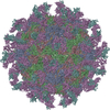
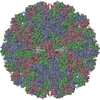
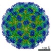
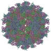
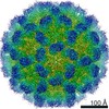
- PDB-1qgc: STRUCTURE OF THE COMPLEX OF A FAB FRAGMENT OF A NEUTRALIZING ANTI... -
+ Open data

- Basic information
| Entry | Database: PDB / ID: 1qgc | ||||||
|---|---|---|---|---|---|---|---|
| Title | STRUCTURE OF THE COMPLEX OF A FAB FRAGMENT OF A NEUTRALIZING ANTIBODY WITH FOOT AND MOUTH DISEASE VIRUS | ||||||
 Components Components |
| ||||||
 Keywords Keywords | Virus/Immune system / VIRUS-ANTIBODY COMPLEX / Icosahedral virus / Virus-Immune system COMPLEX | ||||||
| Function / homology |  Function and homology information Function and homology information: /  L-peptidase / modulation by virus of host chromatin organization / positive stranded viral RNA replication / RNA-protein covalent cross-linking / : / IgG immunoglobulin complex / picornain 3C / ribonucleoside triphosphate phosphatase activity / B cell differentiation ...: / L-peptidase / modulation by virus of host chromatin organization / positive stranded viral RNA replication / RNA-protein covalent cross-linking / : / IgG immunoglobulin complex / picornain 3C / ribonucleoside triphosphate phosphatase activity / B cell differentiation / T=pseudo3 icosahedral viral capsid / host cell cytoplasmic vesicle membrane / : / nucleoside-triphosphate phosphatase / protein complex oligomerization / regulation of translation / monoatomic ion channel activity / clathrin-dependent endocytosis of virus by host cell / RNA helicase activity / viral protein processing / induction by virus of host autophagy / RNA-directed RNA polymerase / viral RNA genome replication / cysteine-type endopeptidase activity / RNA-dependent RNA polymerase activity / DNA-templated transcription / structural molecule activity / virion attachment to host cell / proteolysis / RNA binding / extracellular region / ATP binding / membrane / plasma membrane L-peptidase / modulation by virus of host chromatin organization / positive stranded viral RNA replication / RNA-protein covalent cross-linking / : / IgG immunoglobulin complex / picornain 3C / ribonucleoside triphosphate phosphatase activity / B cell differentiation ...: / L-peptidase / modulation by virus of host chromatin organization / positive stranded viral RNA replication / RNA-protein covalent cross-linking / : / IgG immunoglobulin complex / picornain 3C / ribonucleoside triphosphate phosphatase activity / B cell differentiation / T=pseudo3 icosahedral viral capsid / host cell cytoplasmic vesicle membrane / : / nucleoside-triphosphate phosphatase / protein complex oligomerization / regulation of translation / monoatomic ion channel activity / clathrin-dependent endocytosis of virus by host cell / RNA helicase activity / viral protein processing / induction by virus of host autophagy / RNA-directed RNA polymerase / viral RNA genome replication / cysteine-type endopeptidase activity / RNA-dependent RNA polymerase activity / DNA-templated transcription / structural molecule activity / virion attachment to host cell / proteolysis / RNA binding / extracellular region / ATP binding / membrane / plasma membraneSimilarity search - Function | ||||||
| Biological species |  Foot-and-mouth disease virus - type C Foot-and-mouth disease virus - type C Mus musculus (house mouse) Mus musculus (house mouse) | ||||||
| Method | ELECTRON MICROSCOPY / single particle reconstruction / cryo EM / Resolution: 30 Å | ||||||
 Authors Authors | Fita, I. | ||||||
 Citation Citation | Journal: EMBO J / Year: 1997 Title: Structure of the complex of an Fab fragment of a neutralizing antibody with foot-and-mouth disease virus: positioning of a highly mobile antigenic loop. Authors: E A Hewat / N Verdaguer / I Fita / W Blakemore / S Brookes / A King / J Newman / E Domingo / M G Mateu / D I Stuart /  Abstract: Data from cryo-electron microscopy and X-ray crystallography have been combined to study the interactions of foot-and-mouth disease virus serotype C (FMDV-C) with a strongly neutralizing monoclonal ...Data from cryo-electron microscopy and X-ray crystallography have been combined to study the interactions of foot-and-mouth disease virus serotype C (FMDV-C) with a strongly neutralizing monoclonal antibody (mAb) SD6. The mAb SD6 binds to the long flexible GH-loop of viral protein 1 (VP1) which also binds to an integrin receptor. The structure of the virus-Fab complex was determined to 30 A resolution using cryo-electron microscopy and image analysis. The known structure of FMDV-C, and of the SD6 Fab co-crystallized with a synthetic peptide corresponding to the GH-loop of VP1, were fitted to the cryo-electron microscope density map. The SD6 Fab is seen to project almost radially from the viral surface in an orientation which is only compatible with monovalent binding of the mAb. Even taking into account the mAb hinge and elbow flexibility, it is not possible to model bivalent binding without severely distorting the Fabs. The bound GH-loop is essentially in what has previously been termed the 'up' position in the best fit Fab orientation. The SD6 Fab interacts almost exclusively with the GH-loop of VP1, making very few other contacts with the viral capsid. The position and orientation of the SD6 Fab bound to FMDV-C is in accord with previous immunogenic data. | ||||||
| History |
|
- Structure visualization
| Movie |
Movie viewer |
|---|---|
| Structure viewer | Molecule: MolmilJmol/JSmol |
- Downloads & links
-Download
| PDBx/mmCIF format | 1qgc.cif.gz | 214.4 KB | Display | PDBx/mmCIF format |
|---|---|---|---|---|
| PDB format | pdb1qgc.ent.gz | 164.6 KB | Display | PDB format |
| PDBx/mmJSON format | 1qgc.json.gz | Tree view | PDBx/mmJSON format | |
| Others |  Other downloads Other downloads |
-Validation report
| Arichive directory | https://data.pdbj.org/pub/pdb/validation_reports/qg/1qgcftp://data.pdbj.org/pub/pdb/validation_reports/qg/1qgc | HTTPS FTP |
|---|
-Related structure data
| Similar structure data |
|---|


-Links
 PDBj
PDBj


- Assembly
| Deposited unit | 
|
|---|---|
| 1 | x 60
|
| 2 |
|
| 3 | x 5
|
| 4 | x 6
|
| 5 | 
|
| Symmetry | Point symmetry: (Hermann–Mauguin notation: 532 / Schoenflies symbol: I (icosahedral)) |
-Components
-PROTEIN (VIRUS CAPSID PROTEIN ... , 3 types, 3 molecules 123
| #1: Protein | Mass: 22667.582 Da / Num. of mol.: 1 / Source method: isolated from a natural source / Source: (natural) Foot-and-mouth disease virus - type C / Genus: Aphthovirus / Species: Foot-and-mouth disease virus / Strain: SEROTYPE C / References: UniProt: Q9QCE2, UniProt: P03311*PLUS |
|---|---|
| #2: Protein | Mass: 24297.367 Da / Num. of mol.: 1 / Source method: isolated from a natural source / Source: (natural) Foot-and-mouth disease virus - type C / Genus: Aphthovirus / Species: Foot-and-mouth disease virus / Strain: SEROTYPE C / References: UniProt: Q9QCE2, UniProt: P03311*PLUS |
| #3: Protein | Mass: 24020.875 Da / Num. of mol.: 1 / Source method: isolated from a natural source / Source: (natural) Foot-and-mouth disease virus - type C / Genus: Aphthovirus / Species: Foot-and-mouth disease virus / Strain: SEROTYPE C / References: UniProt: P15072, UniProt: P03311*PLUS |
-Protein/peptide , 1 types, 1 molecules 5
| #6: Protein/peptide | Mass: 2463.722 Da / Num. of mol.: 1 / Fragment: RESIDUES 133-156 / Source method: isolated from a natural source / Source: (natural) Foot-and-mouth disease virus - type C / Genus: Aphthovirus / Species: Foot-and-mouth disease virus / Strain: SEROTYPE C / References: UniProt: P03311*PLUS |
|---|
-Antibody , 2 types, 2 molecules 4A
| #4: Antibody | Mass: 24013.379 Da / Num. of mol.: 1 / Fragment: FAB / Source method: isolated from a natural source / Source: (natural) Mus musculus (house mouse) / References: UniProt: P01837*PLUS |
|---|---|
| #5: Antibody | Mass: 23442.508 Da / Num. of mol.: 1 / Fragment: FAB / Source method: isolated from a natural source / Source: (natural) Mus musculus (house mouse) |
-Experimental details
-Experiment
| Experiment | Method: ELECTRON MICROSCOPY |
|---|---|
| EM experiment | Aggregation state: PARTICLE / 3D reconstruction method: single particle reconstruction |
- Sample preparation
| Component | Name: FAB FRAGMENT OF A NEUTRALIZING ANTIBODY WITH FOOT AND MOUTH DISEASE VIRUS Type: COMPLEX | |||||||||||||||
|---|---|---|---|---|---|---|---|---|---|---|---|---|---|---|---|---|
| Specimen | Embedding applied: NO / Shadowing applied: NO / Staining applied: NO / Vitrification applied: YES | |||||||||||||||
| Specimen support | Film material: HOLEY CARBON / Grid mesh size: 400 divisions/in. | |||||||||||||||
| Crystal grow | *PLUS Method: vapor diffusion | |||||||||||||||
| Components of the solutions | *PLUS
|
- Electron microscopy imaging
| Microscopy | Model: JEOL 2000EXII |
|---|---|
| Electron gun | Accelerating voltage: 100 kV / Illumination mode: FLOOD BEAM |
| Electron lens | Mode: BRIGHT FIELDBright-field microscopy / Nominal magnification: 30000 X / Nominal defocus max: 3000 nm / Nominal defocus min: 1800 nm |
| Image recording | Electron dose: 20 e/Å2 / Film or detector model: KODAK SO-163 FILM |
- Processing
| EM software |
| ||||||||||||||||
|---|---|---|---|---|---|---|---|---|---|---|---|---|---|---|---|---|---|
| Symmetry | Point symmetry: I (icosahedral) | ||||||||||||||||
| 3D reconstruction | Symmetry type: POINT | ||||||||||||||||
| Atomic model building | Space: REAL Details: DETAILS--THE ATOMIC MODEL WAS GENERATED USING THE 3D MAP DETERMINED BY CRYO-ELECTRON MICROSCOPY. X-RAY ATOMIC STRUCTURES WERE AVAILABLE FOR THE INTACT PARTICLE OF FMDV-C AND THE SD6 FAB CO- ...Details: DETAILS--THE ATOMIC MODEL WAS GENERATED USING THE 3D MAP DETERMINED BY CRYO-ELECTRON MICROSCOPY. X-RAY ATOMIC STRUCTURES WERE AVAILABLE FOR THE INTACT PARTICLE OF FMDV-C AND THE SD6 FAB CO-CRYSTALLIZED WITH A SYNTHETIC PEPTIDE CORRESPONDING TO THE DOMINANT ANTIGENIC LOOP, THE GH LOOP, FROM THE VIRAL CAPSIDE PROTEIN VP1. THE ATOMIC MODEL WAS OBTAINED BY DOCKING THE TWO CRYSTALLOGRAPHIC STRUCTURES IN THE RECONSTRUCTED EM MAP. FITTING WAS DONE SIMULTANEOUSLY IN REAL AND RECIPROCAL SPACE. IN REAL SPACE THE CALCULATED ELECTRON DENSITY FOR THE STARTING MODELS WERE FITTED INTO THE CRYO-EM DENSITY BY A STEEPEST DESCENT PROCEDURE WHICH OPTIMIZED THE LINEAR CORRELATION COEFFICIENT BETWEEN THE TWO DISTRIBUTION (GAP- J.GRIMES,D.STUART, UNPUBLISHED) FOR THE RECIPROCAL SPACE FITTING THE STRUCTURE FACTORS CORRESPONDING TO THE CRYO-EM DENSITY OF THE COMPLEX WERE CALCULATED BY INVERSE FOURIER TRANSFORMATIONS AND THESE FACTORS AND PHASES WERE USED TO REFINE THE X-RAY STRUCTURES, BY RIGID BODY MINIMIZATION, USING X-PLOR (BRUNGER, ET.AL., 1993, YALE UNIVERSITY) | ||||||||||||||||
| Atomic model building | PDB-ID: 1FMD Accession code: 1FMD / Source name: PDB / Type: experimental model | ||||||||||||||||
| Refinement | Highest resolution: 30 Å | ||||||||||||||||
| Refinement step | Cycle: LAST / Highest resolution: 30 Å
|

