 ムービー
ムービー コントローラー


+ データを開く

- 基本情報
基本情報
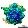
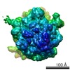
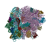
| 登録情報 | データベース: PDB / ID: 1pn7 | ||||||
|---|---|---|---|---|---|---|---|
| タイトル | Coordinates of S12, L11 proteins and P-tRNA, from the 70S X-ray structure aligned to the 70S Cryo-EM map of E.coli ribosome | ||||||
 要素 要素 |
| ||||||
 キーワード キーワード | RNA binding protein/RNA /  ribosomal protein / tRNA binding protein / tRNA (転移RNA) / RNA binding protein-RNA COMPLEX ribosomal protein / tRNA binding protein / tRNA (転移RNA) / RNA binding protein-RNA COMPLEX | ||||||
| 機能・相同性 |  機能・相同性情報 機能・相同性情報large ribosomal subunit rRNA binding / small ribosomal subunit / cytosolic large ribosomal subunit / tRNA binding / rRNA binding / structural constituent of ribosome / 翻訳 (生物学)類似検索 - 分子機能 | ||||||
| 生物種 |   Thermus thermophilus (サーマス・サーモフィルス)Thermotoga maritima (テルモトガ・マリティマ) Thermus thermophilus (サーマス・サーモフィルス)Thermotoga maritima (テルモトガ・マリティマ) | ||||||
| 手法 | 電子顕微鏡法 / 単粒子再構成法 / クライオ電子顕微鏡法 / 解像度: 10.8 Å | ||||||
 データ登録者 データ登録者 | Valle, M. / Zavialov, A. / Sengupta, J. / Rawat, U. / Ehrenberg, M. / Frank, J. | ||||||
 引用 引用 | ジャーナル: Cell / 年: 2003 タイトル: Locking and unlocking of ribosomal motions. 著者: Mikel Valle / Andrey Zavialov / Jayati Sengupta / Urmila Rawat / Måns Ehrenberg / Joachim Frank /  要旨: During the ribosomal translocation, the binding of elongation factor G (EF-G) to the pretranslocational ribosome leads to a ratchet-like rotation of the 30S subunit relative to the 50S subunit in the ...During the ribosomal translocation, the binding of elongation factor G (EF-G) to the pretranslocational ribosome leads to a ratchet-like rotation of the 30S subunit relative to the 50S subunit in the direction of the mRNA movement. By means of cryo-electron microscopy we observe that this rotation is accompanied by a 20 A movement of the L1 stalk of the 50S subunit, implying that this region is involved in the translocation of deacylated tRNAs from the P to the E site. These ribosomal motions can occur only when the P-site tRNA is deacylated. Prior to peptidyl-transfer to the A-site tRNA or peptide removal, the presence of the charged P-site tRNA locks the ribosome and prohibits both of these motions. | ||||||
| 履歴 |
| ||||||
| Remark 999 | The structure contains C alpha atoms only |
- 構造の表示
構造の表示
| ムービー |
ムービービューア |
|---|---|
| 構造ビューア | 分子: MolmilJmol/JSmol |
- ダウンロードとリンク
-ダウンロード
| PDBx/mmCIF形式 | 1pn7.cif.gz | 23.1 KB | 表示 | PDBx/mmCIF形式 |
|---|---|---|---|---|
| PDB形式 | pdb1pn7.ent.gz | 9.2 KB | 表示 | PDB形式 |
| PDBx/mmJSON形式 | 1pn7.json.gz | ツリー表示 | PDBx/mmJSON形式 | |
| その他 |  その他のダウンロード その他のダウンロード |
-検証レポート
| アーカイブディレクトリ | https://data.pdbj.org/pub/pdb/validation_reports/pn/1pn7ftp://data.pdbj.org/pub/pdb/validation_reports/pn/1pn7 | HTTPS FTP |
|---|
-関連構造データ
-リンク
 PDBj
PDBj


- 集合体
集合体
| 登録構造単位 | 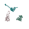
|
|---|---|
| 1 |
|
-要素
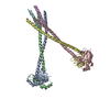
| #1: RNA鎖 | 分子量: 20016.959 Da / 分子数: 1 / 由来タイプ: 合成 |
|---|---|
| #2: タンパク質 | / 座標モデル: Cα原子のみ 分子量: 13804.311 Da / 分子数: 1 / 由来タイプ: 天然 由来: (天然) Thermus thermophilus (サーマス・サーモフィルス)参照: UniProt: Q5SHN3 |
| #3: タンパク質 | / 座標モデル: Cα原子のみ 分子量: 14294.913 Da / 分子数: 1 / 由来タイプ: 天然 由来: (天然) Thermotoga maritima (テルモトガ・マリティマ)参照: UniProt: P29395 |
-実験情報
-実験
| 実験 | 手法: 電子顕微鏡法 |
|---|---|
| EM実験 | 試料の集合状態: PARTICLE / 3次元再構成法: 単粒子再構成法 |
- 試料調製
試料調製
| 構成要素 |
| ||||||||||||||||||||
|---|---|---|---|---|---|---|---|---|---|---|---|---|---|---|---|---|---|---|---|---|---|
| 緩衝液 | pH: 7.5 | ||||||||||||||||||||
| 試料 | 濃度: 32 mg/ml / 包埋: NO / シャドウイング: NO / 染色: NO / 凍結: YES | ||||||||||||||||||||
| 試料支持 | 詳細: Quantifoil holley-carbon film grids | ||||||||||||||||||||
| 急速凍結 | 凍結剤: ETHANE / 詳細: Rapid-freezing in liquid ethane | ||||||||||||||||||||
| 結晶化 | *PLUS 手法: 電子顕微鏡法 / 詳細: electron microscopy |
- 電子顕微鏡撮影
| 実験機器 |  モデル: Tecnai F20 / 画像提供: FEI Company |
|---|---|
| 顕微鏡 | モデル: FEI TECNAI F20 / 日付: 2001年6月1日 |
| 電子銃 | 電子線源: FIELD EMISSION GUN / 加速電圧: 200 kV / 照射モード: FLOOD BEAM |
| 電子レンズ | モード: BRIGHT FIELDBright-field microscopy / 倍率(公称値): 50000 X / 倍率(補正後): 49696 X / 最大 デフォーカス(公称値): 4000 nm / 最小 デフォーカス(公称値): 1500 nm / Cs: 2 mm |
| 試料ホルダ | 温度: 93 K / 傾斜角・最大: 0 ° / 傾斜角・最小: 0 ° |
| 撮影 | 電子線照射量: 20 e/Å2 / フィルム・検出器のモデル: KODAK SO-163 FILM |
- 解析
解析
| CTF補正 | 詳細: CTF correction of 3D-maps by Wiener filtration | |||||||||||||||||||||
|---|---|---|---|---|---|---|---|---|---|---|---|---|---|---|---|---|---|---|---|---|---|---|
| 対称性 | 点対称性: C1 (非対称) | |||||||||||||||||||||
| 3次元再構成 | 手法: 3D projection matching; conjugate gradients with regularization 解像度: 10.8 Å / ピクセルサイズ(実測値): 2.82 Å / 倍率補正: TMV 詳細: SPIDER package. Crystal Structure of Thermus Thermophilus 70S ribosome 対称性のタイプ: POINT | |||||||||||||||||||||
| 原子モデル構築 | プロトコル: OTHER / 空間: REAL / 詳細: METHOD--Manual fitting in O | |||||||||||||||||||||
| 原子モデル構築 |
| |||||||||||||||||||||
| 精密化ステップ | サイクル: LAST
|

