 Movie
Movie Controller

[English] 日本語
 Yorodumi
- PDB-1pn7: Coordinates of S12, L11 proteins and P-tRNA, from the 70S X-ray s... -
+ Open data

- Basic information
| Entry | Database: PDB / ID: 1pn7 | ||||||
|---|---|---|---|---|---|---|---|
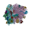
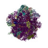
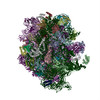
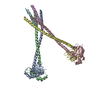
| Title | Coordinates of S12, L11 proteins and P-tRNA, from the 70S X-ray structure aligned to the 70S Cryo-EM map of E.coli ribosome | ||||||
 Components Components |
| ||||||
 Keywords Keywords | RNA binding protein/RNA /  ribosomal protein / tRNA binding protein / tRNA / RNA binding protein-RNA COMPLEX ribosomal protein / tRNA binding protein / tRNA / RNA binding protein-RNA COMPLEX | ||||||
| Function / homology |  Function and homology information Function and homology informationlarge ribosomal subunit rRNA binding / small ribosomal subunit / cytosolic large ribosomal subunit / tRNA binding / rRNA binding / structural constituent of ribosome / translationSimilarity search - Function | ||||||
| Biological species |   Thermus thermophilus (bacteria)Thermotoga maritima (bacteria) Thermus thermophilus (bacteria)Thermotoga maritima (bacteria) | ||||||
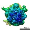
| Method | ELECTRON MICROSCOPY / single particle reconstruction / cryo EM / Resolution: 10.8 Å | ||||||
 Authors Authors | Valle, M. / Zavialov, A. / Sengupta, J. / Rawat, U. / Ehrenberg, M. / Frank, J. | ||||||
 Citation Citation | Journal: Cell / Year: 2003 Title: Locking and unlocking of ribosomal motions. Authors: Mikel Valle / Andrey Zavialov / Jayati Sengupta / Urmila Rawat / Måns Ehrenberg / Joachim Frank /  Abstract: During the ribosomal translocation, the binding of elongation factor G (EF-G) to the pretranslocational ribosome leads to a ratchet-like rotation of the 30S subunit relative to the 50S subunit in the ...During the ribosomal translocation, the binding of elongation factor G (EF-G) to the pretranslocational ribosome leads to a ratchet-like rotation of the 30S subunit relative to the 50S subunit in the direction of the mRNA movement. By means of cryo-electron microscopy we observe that this rotation is accompanied by a 20 A movement of the L1 stalk of the 50S subunit, implying that this region is involved in the translocation of deacylated tRNAs from the P to the E site. These ribosomal motions can occur only when the P-site tRNA is deacylated. Prior to peptidyl-transfer to the A-site tRNA or peptide removal, the presence of the charged P-site tRNA locks the ribosome and prohibits both of these motions. | ||||||
| History |
| ||||||
| Remark 999 | The structure contains C alpha atoms only |
- Structure visualization
| Movie |
Movie viewer |
|---|---|
| Structure viewer | Molecule: MolmilJmol/JSmol |
- Downloads & links
-Download
| PDBx/mmCIF format | 1pn7.cif.gz | 23.1 KB | Display | PDBx/mmCIF format |
|---|---|---|---|---|
| PDB format | pdb1pn7.ent.gz | 9.2 KB | Display | PDB format |
| PDBx/mmJSON format | 1pn7.json.gz | Tree view | PDBx/mmJSON format | |
| Others |  Other downloads Other downloads |
-Validation report
| Arichive directory | https://data.pdbj.org/pub/pdb/validation_reports/pn/1pn7ftp://data.pdbj.org/pub/pdb/validation_reports/pn/1pn7 | HTTPS FTP |
|---|
-Related structure data
| Related structure data |  1362MC  1363MC  1364MC  1365MC 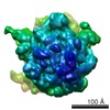 1366MC 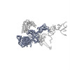 1pn6C 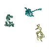 1pn8C C: citing same article ( M: map data used to model this data |
|---|---|
| Similar structure data |
-Links
 PDBj
PDBj


- Assembly
| Deposited unit | 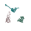
|
|---|---|
| 1 |
|
-Components
| #1: RNA chain | Mass: 20016.959 Da / Num. of mol.: 1 / Source method: obtained synthetically |
|---|---|
| #2: Protein | / Coordinate model: Cα atoms only Mass: 13804.311 Da / Num. of mol.: 1 / Source method: isolated from a natural source / Source: (natural) Thermus thermophilus (bacteria) / References: UniProt: Q5SHN3 |
| #3: Protein | / Coordinate model: Cα atoms only Mass: 14294.913 Da / Num. of mol.: 1 / Source method: isolated from a natural source / Source: (natural) Thermotoga maritima (bacteria) / References: UniProt: P29395 |
-Experimental details
-Experiment
| Experiment | Method: ELECTRON MICROSCOPY |
|---|---|
| EM experiment | Aggregation state: PARTICLE / 3D reconstruction method: single particle reconstruction |
- Sample preparation
| Component |
| ||||||||||||||||||||
|---|---|---|---|---|---|---|---|---|---|---|---|---|---|---|---|---|---|---|---|---|---|
| Buffer solution | pH: 7.5 | ||||||||||||||||||||
| Specimen | Conc.: 32 mg/ml / Embedding applied: NO / Shadowing applied: NO / Staining applied: NO / Vitrification applied: YES | ||||||||||||||||||||
| Specimen support | Details: Quantifoil holley-carbon film grids | ||||||||||||||||||||
| Vitrification | Cryogen name: ETHANE / Details: Rapid-freezing in liquid ethane | ||||||||||||||||||||
| Crystal grow | *PLUS Method: electron microscopy / Details: electron microscopy |
- Electron microscopy imaging
| Experimental equipment |  Model: Tecnai F20 / Image courtesy: FEI Company |
|---|---|
| Microscopy | Model: FEI TECNAI F20 / Date: Jun 1, 2001 |
| Electron gun | Electron source: FIELD EMISSION GUN / Accelerating voltage: 200 kV / Illumination mode: FLOOD BEAM |
| Electron lens | Mode: BRIGHT FIELDBright-field microscopy / Nominal magnification: 50000 X / Calibrated magnification: 49696 X / Nominal defocus max: 4000 nm / Nominal defocus min: 1500 nm / Cs: 2 mm |
| Specimen holder | Temperature: 93 K / Tilt angle max: 0 ° / Tilt angle min: 0 ° |
| Image recording | Electron dose: 20 e/Å2 / Film or detector model: KODAK SO-163 FILM |
- Processing
| CTF correction | Details: CTF correction of 3D-maps by Wiener filtration | |||||||||||||||||||||
|---|---|---|---|---|---|---|---|---|---|---|---|---|---|---|---|---|---|---|---|---|---|---|
| Symmetry | Point symmetry: C1 (asymmetric) | |||||||||||||||||||||
| 3D reconstruction | Method: 3D projection matching; conjugate gradients with regularization Resolution: 10.8 Å / Actual pixel size: 2.82 Å / Magnification calibration: TMV Details: SPIDER package. Crystal Structure of Thermus Thermophilus 70S ribosome Symmetry type: POINT | |||||||||||||||||||||
| Atomic model building | Protocol: OTHER / Space: REAL / Details: METHOD--Manual fitting in O | |||||||||||||||||||||
| Atomic model building |
| |||||||||||||||||||||
| Refinement step | Cycle: LAST
|

