 ムービー
ムービー コントローラー


+ データを開く

- 基本情報
基本情報
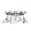
| 登録情報 | データベース: PDB / ID: 1pdi | ||||||
|---|---|---|---|---|---|---|---|
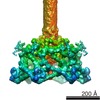
| タイトル | Fitting of the C-terminal part of the short tail fibers into the cryo-EM reconstruction of T4 baseplate | ||||||
 要素 要素 | Short tail fiber protein | ||||||
 キーワード キーワード |  STRUCTURAL PROTEIN (タンパク質) STRUCTURAL PROTEIN (タンパク質) | ||||||
| 機能・相同性 |  機能・相同性情報 機能・相同性情報virus tail, baseplate / virus tail, fiber / entry receptor-mediated virion attachment to host cell / symbiont entry into host cell / metal ion binding類似検索 - 分子機能 | ||||||
| 生物種 |  Enterobacteria phage T4 (ファージ) Enterobacteria phage T4 (ファージ) | ||||||
| 手法 | 電子顕微鏡法 / 単粒子再構成法 / クライオ電子顕微鏡法 / 解像度: 12 Å | ||||||
 データ登録者 データ登録者 | Kostyuchenko, V.A. / Leiman, P.G. / Chipman, P.R. / Kanamaru, S. / van Raaij, M.J. / Arisaka, F. / Mesyanzhinov, V.V. / Rossmann, M.G. | ||||||
 引用 引用 | ジャーナル: Nat Struct Biol / 年: 2003 タイトル: Three-dimensional structure of bacteriophage T4 baseplate. 著者: Victor A Kostyuchenko / Petr G Leiman / Paul R Chipman / Shuji Kanamaru / Mark J van Raaij / Fumio Arisaka / Vadim V Mesyanzhinov / Michael G Rossmann /  要旨: The baseplate of bacteriophage T4 is a multiprotein molecular machine that controls host cell recognition, attachment, tail sheath contraction and viral DNA ejection. We report here the three- ...The baseplate of bacteriophage T4 is a multiprotein molecular machine that controls host cell recognition, attachment, tail sheath contraction and viral DNA ejection. We report here the three-dimensional structure of the baseplate-tail tube complex determined to a resolution of 12 A by cryoelectron microscopy. The baseplate has a six-fold symmetric, dome-like structure approximately 520 A in diameter and approximately 270 A long, assembled around a central hub. A 940 A-long and 96 A-diameter tail tube, coaxial with the hub, is connected to the top of the baseplate. At the center of the dome is a needle-like structure that was previously identified as a cell puncturing device. We have identified the locations of six proteins with known atomic structures, and established the position and shape of several other baseplate proteins. The baseplate structure suggests a mechanism of baseplate triggering and structural transition during the initial stages of T4 infection. #1: ジャーナル: J.Mol.Biol. / 年: 2001タイトル: Crystal Structure of a Heat-and Protease-Stable Part of the Bacteriophage T4 Short Tail Fibre 著者: van Raaij, M.J. / Schoehn, G. / Burda, M.R. / Miller, S. #2: ジャーナル: To be publishedタイトル: Structure of the receptor-binding domain of the bacteriophage T4 short tail fibre 著者: Thomassen, E. / Gielen, G. / Schuetz, M. / Miller, S. / van Raaij, M.J. | ||||||
| 履歴 |
| ||||||
| Remark 999 | SEQUENCE The differences between the database reference sequence and the deposited sequence arise ...SEQUENCE The differences between the database reference sequence and the deposited sequence arise because the source of the fitted model is bacteriophage T4alc7, whereas the protein used in the structural solution is from bacteriophage T4D. The sequence of the protein from T4D has not yet been deposited. Only coordinates for CA atoms were submitted. | ||||||
| Remark 300 | BIOMOLECULE: 1 THIS ENTRY CONTAINS A PORTION OF THE BIOLOGICALLY SIGNIFICANT MULTIMER. ASSEMBLY ...BIOMOLECULE: 1 THIS ENTRY CONTAINS A PORTION OF THE BIOLOGICALLY SIGNIFICANT MULTIMER. ASSEMBLY COMPONENTS COM_ID: 1 NAME:GP12 IPR_ID: NULL GO_ID: NULL OTHER_DETAILS: TRIMER |
- 構造の表示
構造の表示
| ムービー |
ムービービューア |
|---|---|
| 構造ビューア | 分子: MolmilJmol/JSmol |
- ダウンロードとリンク
-ダウンロード
| PDBx/mmCIF形式 | 1pdi.cif.gz | 138.1 KB | 表示 | PDBx/mmCIF形式 |
|---|---|---|---|---|
| PDB形式 | pdb1pdi.ent.gz | 95.1 KB | 表示 | PDB形式 |
| PDBx/mmJSON形式 | 1pdi.json.gz | ツリー表示 | PDBx/mmJSON形式 | |
| その他 |  その他のダウンロード その他のダウンロード |
-検証レポート
| アーカイブディレクトリ | https://data.pdbj.org/pub/pdb/validation_reports/pd/1pdiftp://data.pdbj.org/pub/pdb/validation_reports/pd/1pdi | HTTPS FTP |
|---|
-関連構造データ


-リンク
 PDBj
PDBj- 集合体
集合体
| 登録構造単位 | 
|
|---|---|
| 1 |
|
| 対称性 | 点対称性: (シェーンフリース記号: C6 (6回回転対称)) |
-要素
| #1: タンパク質 | 分子量: 29856.020 Da / 分子数: 18 / 由来タイプ: 天然 / 由来: (天然) Enterobacteria phage T4 (ファージ) / 属: T4-like viruses / 生物種: Enterobacteria phage T4 sensu lato / 株: T4D / 参照: UniProt: P10930 |
|---|
-実験情報
-実験
| 実験 | 手法: 電子顕微鏡法 |
|---|---|
| EM実験 | 試料の集合状態: PARTICLE / 3次元再構成法: 単粒子再構成法 |
- 試料調製
試料調製
| 構成要素 |
| |||||||||||||||
|---|---|---|---|---|---|---|---|---|---|---|---|---|---|---|---|---|
| 緩衝液 | 名称: water水 / pH: 7 / 詳細: water | |||||||||||||||
| 試料 | 濃度: 5 mg/ml / 包埋: NO / シャドウイング: NO / 染色: NO / 凍結: YES | |||||||||||||||
| 試料支持 | 詳細: holey carbon | |||||||||||||||
| 急速凍結 | 凍結剤: ETHANE / 詳細: ethane vitrification | |||||||||||||||
| 結晶化 | *PLUS 手法: 電子顕微鏡法 |
- 電子顕微鏡撮影
| 顕微鏡 | モデル: FEI/PHILIPS CM300FEG/T / 日付: 2001年1月30日 |
|---|---|
| 電子銃 | 電子線源: FIELD EMISSION GUN / 加速電圧: 300 kV / 照射モード: FLOOD BEAM |
| 電子レンズ | モード: BRIGHT FIELDBright-field microscopy / 倍率(公称値): 45000 X / 倍率(補正後): 47000 X / 最大 デフォーカス(公称値): 5000 nm / 最小 デフォーカス(公称値): 1200 nm / Cs: 2 mm |
| 試料ホルダ | 温度: 70 K / 傾斜角・最大: 0 ° / 傾斜角・最小: 0 ° |
| 撮影 | 電子線照射量: 25 e/Å2 / フィルム・検出器のモデル: KODAK SO-163 FILM |
- 解析
解析
| EMソフトウェア |
| ||||||||||||
|---|---|---|---|---|---|---|---|---|---|---|---|---|---|
| CTF補正 | 詳細: CTF correction of individual particles with Wiener filtering | ||||||||||||
| 対称性 | 点対称性: C6 (6回回転対称) | ||||||||||||
| 3次元再構成 | 手法: model-based projection matching / 解像度: 12 Å / 粒子像の数: 945 / ピクセルサイズ(公称値): 3.11 Å / ピクセルサイズ(実測値): 2.98 Å / 倍率補正: TMV particles images 詳細: modifed version of program SPIDER was used for the reconstruction 対称性のタイプ: POINT | ||||||||||||
| 原子モデル構築 | プロトコル: OTHER / 空間: REAL / Target criteria: Correlation Coefficient maximization / 詳細: REFINEMENT PROTOCOL--Laplacian transform real space | ||||||||||||
| 原子モデル構築 | PDB-ID: 1OCY Accession code: 1OCY / Source name: PDB / タイプ: experimental model | ||||||||||||
| 精密化ステップ | サイクル: LAST
|
