 Movie
Movie Controller

[English] 日本語
 Yorodumi
- PDB-1pdi: Fitting of the C-terminal part of the short tail fibers into the ... -
+ Open data

- Basic information
| Entry | Database: PDB / ID: 1pdi | ||||||
|---|---|---|---|---|---|---|---|
| Title | Fitting of the C-terminal part of the short tail fibers into the cryo-EM reconstruction of T4 baseplate | ||||||
 Components Components | Short tail fiber protein | ||||||
 Keywords Keywords | STRUCTURAL PROTEIN | ||||||
| Function / homology |  Function and homology information Function and homology informationvirus tail, baseplate / virus tail, fiber / entry receptor-mediated virion attachment to host cell / symbiont entry into host cell / metal ion binding Similarity search - Function | ||||||
| Biological species |  Enterobacteria phage T4 (virus) Enterobacteria phage T4 (virus) | ||||||
| Method | ELECTRON MICROSCOPY / single particle reconstruction / cryo EM / Resolution: 12 Å | ||||||
 Authors Authors | Kostyuchenko, V.A. / Leiman, P.G. / Chipman, P.R. / Kanamaru, S. / van Raaij, M.J. / Arisaka, F. / Mesyanzhinov, V.V. / Rossmann, M.G. | ||||||
 Citation Citation | Journal: Nat Struct Biol / Year: 2003 Title: Three-dimensional structure of bacteriophage T4 baseplate. Authors: Victor A Kostyuchenko / Petr G Leiman / Paul R Chipman / Shuji Kanamaru / Mark J van Raaij / Fumio Arisaka / Vadim V Mesyanzhinov / Michael G Rossmann /  Abstract: The baseplate of bacteriophage T4 is a multiprotein molecular machine that controls host cell recognition, attachment, tail sheath contraction and viral DNA ejection. We report here the three- ...The baseplate of bacteriophage T4 is a multiprotein molecular machine that controls host cell recognition, attachment, tail sheath contraction and viral DNA ejection. We report here the three-dimensional structure of the baseplate-tail tube complex determined to a resolution of 12 A by cryoelectron microscopy. The baseplate has a six-fold symmetric, dome-like structure approximately 520 A in diameter and approximately 270 A long, assembled around a central hub. A 940 A-long and 96 A-diameter tail tube, coaxial with the hub, is connected to the top of the baseplate. At the center of the dome is a needle-like structure that was previously identified as a cell puncturing device. We have identified the locations of six proteins with known atomic structures, and established the position and shape of several other baseplate proteins. The baseplate structure suggests a mechanism of baseplate triggering and structural transition during the initial stages of T4 infection. #1: Journal: J.Mol.Biol. / Year: 2001Title: Crystal Structure of a Heat-and Protease-Stable Part of the Bacteriophage T4 Short Tail Fibre Authors: van Raaij, M.J. / Schoehn, G. / Burda, M.R. / Miller, S. #2: Journal: To be publishedTitle: Structure of the receptor-binding domain of the bacteriophage T4 short tail fibre Authors: Thomassen, E. / Gielen, G. / Schuetz, M. / Miller, S. / van Raaij, M.J. | ||||||
| History |
| ||||||
| Remark 999 | SEQUENCE The differences between the database reference sequence and the deposited sequence arise ...SEQUENCE The differences between the database reference sequence and the deposited sequence arise because the source of the fitted model is bacteriophage T4alc7, whereas the protein used in the structural solution is from bacteriophage T4D. The sequence of the protein from T4D has not yet been deposited. Only coordinates for CA atoms were submitted. | ||||||
| Remark 300 | BIOMOLECULE: 1 THIS ENTRY CONTAINS A PORTION OF THE BIOLOGICALLY SIGNIFICANT MULTIMER. ASSEMBLY ...BIOMOLECULE: 1 THIS ENTRY CONTAINS A PORTION OF THE BIOLOGICALLY SIGNIFICANT MULTIMER. ASSEMBLY COMPONENTS COM_ID: 1 NAME:GP12 IPR_ID: NULL GO_ID: NULL OTHER_DETAILS: TRIMER |
- Structure visualization
| Movie |
Movie viewer |
|---|---|
| Structure viewer | Molecule: MolmilJmol/JSmol |
- Downloads & links
-Download
| PDBx/mmCIF format | 1pdi.cif.gz | 138.1 KB | Display | PDBx/mmCIF format |
|---|---|---|---|---|
| PDB format | pdb1pdi.ent.gz | 95.1 KB | Display | PDB format |
| PDBx/mmJSON format | 1pdi.json.gz | Tree view | PDBx/mmJSON format | |
| Others |  Other downloads Other downloads |
-Validation report
| Summary document | 1pdi_validation.pdf.gz | 766.3 KB | Display | wwPDB validaton report |
|---|---|---|---|---|
| Full document | 1pdi_full_validation.pdf.gz | 765.5 KB | Display | |
| Data in XML | 1pdi_validation.xml.gz | 55.6 KB | Display | |
| Data in CIF | 1pdi_validation.cif.gz | 84.7 KB | Display | |
| Arichive directory | https://data.pdbj.org/pub/pdb/validation_reports/pd/1pdiftp://data.pdbj.org/pub/pdb/validation_reports/pd/1pdi | HTTPS FTP |
-Related structure data
| Related structure data | 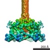 1048MC  1pdfC  1pdjC  1pdlC 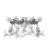 1pdmC  1pdpC M: map data used to model this data C: citing same article ( |
|---|---|
| Similar structure data |


-Links
 PDBj
PDBj- Assembly
| Deposited unit | 
|
|---|---|
| 1 |
|
| Symmetry | Point symmetry: (Schoenflies symbol: C6 (6 fold cyclic)) |
-Components
| #1: Protein | Mass: 29856.020 Da / Num. of mol.: 18 / Source method: isolated from a natural source / Source: (natural) Enterobacteria phage T4 (virus) / Genus: T4-like viruses / Species: Enterobacteria phage T4 sensu lato / Strain: T4D / References: UniProt: P10930 |
|---|
-Experimental details
-Experiment
| Experiment | Method: ELECTRON MICROSCOPY |
|---|---|
| EM experiment | Aggregation state: PARTICLE / 3D reconstruction method: single particle reconstruction |
- Sample preparation
| Component |
| |||||||||||||||
|---|---|---|---|---|---|---|---|---|---|---|---|---|---|---|---|---|
| Buffer solution | Name: water / pH: 7 / Details: water | |||||||||||||||
| Specimen | Conc.: 5 mg/ml / Embedding applied: NO / Shadowing applied: NO / Staining applied: NO / Vitrification applied: YES | |||||||||||||||
| Specimen support | Details: holey carbon | |||||||||||||||
| Vitrification | Cryogen name: ETHANE / Details: ethane vitrification | |||||||||||||||
| Crystal grow | *PLUS Method: electron microscopy |
- Electron microscopy imaging
| Microscopy | Model: FEI/PHILIPS CM300FEG/T / Date: Jan 30, 2001 |
|---|---|
| Electron gun | Electron source:  FIELD EMISSION GUN / Accelerating voltage: 300 kV / Illumination mode: FLOOD BEAM FIELD EMISSION GUN / Accelerating voltage: 300 kV / Illumination mode: FLOOD BEAM |
| Electron lens | Mode: BRIGHT FIELD / Nominal magnification: 45000 X / Calibrated magnification: 47000 X / Nominal defocus max: 5000 nm / Nominal defocus min: 1200 nm / Cs: 2 mm |
| Specimen holder | Temperature: 70 K / Tilt angle max: 0 ° / Tilt angle min: 0 ° |
| Image recording | Electron dose: 25 e/Å2 / Film or detector model: KODAK SO-163 FILM |
- Processing
| EM software |
| ||||||||||||
|---|---|---|---|---|---|---|---|---|---|---|---|---|---|
| CTF correction | Details: CTF correction of individual particles with Wiener filtering | ||||||||||||
| Symmetry | Point symmetry: C6 (6 fold cyclic) | ||||||||||||
| 3D reconstruction | Method: model-based projection matching / Resolution: 12 Å / Num. of particles: 945 / Nominal pixel size: 3.11 Å / Actual pixel size: 2.98 Å / Magnification calibration: TMV particles images Details: modifed version of program SPIDER was used for the reconstruction Symmetry type: POINT | ||||||||||||
| Atomic model building | Protocol: OTHER / Space: REAL / Target criteria: Correlation Coefficient maximization Details: REFINEMENT PROTOCOL--Laplacian transform real space | ||||||||||||
| Atomic model building | PDB-ID: 1OCY Accession code: 1OCY / Source name: PDB / Type: experimental model | ||||||||||||
| Refinement step | Cycle: LAST
|
