 Movie
Movie Controller

[English] 日本語
 Yorodumi
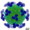
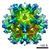
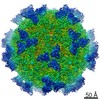
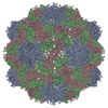
- PDB-1kvp: STRUCTURAL ANALYSIS OF THE SPIROPLASMA VIRUS, SPV4, IMPLICATIONS ... -
+ Open data

- Basic information
| Entry | Database: PDB / ID: 1kvp | ||||||
|---|---|---|---|---|---|---|---|
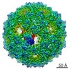
| Title | STRUCTURAL ANALYSIS OF THE SPIROPLASMA VIRUS, SPV4, IMPLICATIONS FOR EVOLUTIONARY VARIATION TO OBTAIN HOST DIVERSITY AMONG THE MICROVIRIDAE, ELECTRON MICROSCOPY, ALPHA CARBONS ONLY | ||||||
 Components Components | SPV4 CAPSID PROTEIN VP1 | ||||||
 Keywords Keywords | VIRUS / BACTERIOPHAGE SPV4 COAT PROTEIN / CHIMERA / Icosahedral virus | ||||||
| Function / homology | Microviridae F protein / Microviridae F protein superfamily / Capsid protein (F protein) / Capsid/spike protein, ssDNA virus / T=1 icosahedral viral capsid / symbiont entry into host cell / structural molecule activity / Capsid protein F Function and homology information Function and homology information | ||||||
| Biological species |  Enterobacteria phage phiX174 (virus) Enterobacteria phage phiX174 (virus) | ||||||
| Method | ELECTRON MICROSCOPY / single particle reconstruction / cryo EM / Resolution: 27 Å | ||||||
 Authors Authors | McKenna, R. | ||||||
 Citation Citation | Journal: Structure / Year: 1998 Title: Structural analysis of the Spiroplasma virus, SpV4: implications for evolutionary variation to obtain host diversity among the Microviridae. Authors: P R Chipman / M Agbandje-McKenna / J Renaudin / T S Baker / R McKenna /  Abstract: BACKGROUND: Spiroplasma virus, SpV4, is a small, non-enveloped virus that infects the helical mollicute Spiroplasma melliferum. SpV4 exhibits several similarities to the Chlamydia phage, Chp1, and ...BACKGROUND: Spiroplasma virus, SpV4, is a small, non-enveloped virus that infects the helical mollicute Spiroplasma melliferum. SpV4 exhibits several similarities to the Chlamydia phage, Chp1, and the Coliphages alpha 3, phi K, G4 and phi X174. All of these viruses are members of the Microviridae. These viruses have isometric capsids with T = 1 icosahedral symmetry, cause lytic infections and are the only icosahedral phages that contain single-stranded circular DNA genomes. The aim of this comparative study on these phages was to understand the role of their capsid proteins during host receptor recognition. RESULTS: The three-dimensional structure of SpV4 was determined to 27 A resolution from images of frozen-hydrated particles. Cryo-electron microscopy (cryo-EM) revealed 20, approximately 54 A long, ...RESULTS: The three-dimensional structure of SpV4 was determined to 27 A resolution from images of frozen-hydrated particles. Cryo-electron microscopy (cryo-EM) revealed 20, approximately 54 A long, 'mushroom-like' protrusions on the surface of the capsid. Each protrusion comprises a trimeric structure that extends radially along the threefold icosahedral axes of the capsid. A 71 amino acid portion of VP1 (the SpV4 capsid protein) was shown, by structural alignment with the atomic structure of the F capsid protein of phi X174, to represent an insertion sequence between the E and F strands of the eight-stranded antiparallel beta-barrel. Secondary structure prediction of this insertion sequence provided the basis for a probable structural motif, consisting of a six-stranded antiparallel beta sheet connected by small turns. Three such motifs form the rigid stable trimeric structures (mushroom-like protrusions) at the threefold axes, with hydrophobic depressions at their distal surface. CONCLUSIONS: Sequence alignment and structural analysis indicate that distinct genera of the Microviridae might have evolved from a common primordial ancestor, with capsid surface variations, such as ...CONCLUSIONS: Sequence alignment and structural analysis indicate that distinct genera of the Microviridae might have evolved from a common primordial ancestor, with capsid surface variations, such as the SpV4 protrusions, resulting from gene fusion events that have enabled diverse host ranges. The hydrophobic nature of the cavity at the distal surface of the SpV4 protrusions suggests that this region may function as the receptor-recognition site during host infection. #1: Journal: Nature / Year: 1992Title: Atomic Structure of Single-Stranded DNA Bacteriophage Phi X174 and its Functional Implications Authors: McKenna, R. / Xia, D. / Willingmann, P. / Ilag, L.L. / Krishnaswamy, S. / Rossmann, M.G. / Olson, N.H. / Baker, T.S. / Incardona, N.L. #2: Journal: Isr.J.Med.Sci. / Year: 1984Title: Characterization of Spiroplasma Virus Group 4 (Sv4) Authors: Renaudin, J. / Pascarel, M.C. / Garnier, M. / Carle, P. / Bove, J.M. | ||||||
| History |
|
- Structure visualization
| Movie |
Movie viewer |
|---|---|
| Structure viewer | Molecule: MolmilJmol/JSmol |
- Downloads & links
-Download
| PDBx/mmCIF format | 1kvp.cif.gz | 31.7 KB | Display | PDBx/mmCIF format |
|---|---|---|---|---|
| PDB format | pdb1kvp.ent.gz | 14.7 KB | Display | PDB format |
| PDBx/mmJSON format | 1kvp.json.gz | Tree view | PDBx/mmJSON format | |
| Others |  Other downloads Other downloads |
-Validation report
| Summary document | 1kvp_validation.pdf.gz | 236.3 KB | Display | wwPDB validaton report |
|---|---|---|---|---|
| Full document | 1kvp_full_validation.pdf.gz | 235.8 KB | Display | |
| Data in XML | 1kvp_validation.xml.gz | 703 B | Display | |
| Data in CIF | 1kvp_validation.cif.gz | 4.8 KB | Display | |
| Arichive directory | https://data.pdbj.org/pub/pdb/validation_reports/kv/1kvpftp://data.pdbj.org/pub/pdb/validation_reports/kv/1kvp | HTTPS FTP |
-Related structure data
| Similar structure data |
|---|
-Links
 PDBj
PDBj


- Assembly
| Deposited unit | 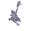
|
|---|---|
| 1 | x 60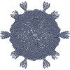
|
| 2 |
|
| 3 | x 5
|
| 4 | x 6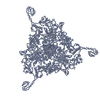
|
| 5 | 
|
| Symmetry | Point symmetry: (Hermann–Mauguin notation: 532 / Schoenflies symbol: I (icosahedral)) |
-Components
| #1: Protein | Mass: 55834.703 Da / Num. of mol.: 1 Source method: isolated from a genetically manipulated source Source: (gene. exp.) Enterobacteria phage phiX174 (virus) / Production host:  Spiroplasma melliferum (bacteria) / References: UniProt: P03641 Spiroplasma melliferum (bacteria) / References: UniProt: P03641 |
|---|
-Experimental details
-Experiment
| Experiment | Method: ELECTRON MICROSCOPY |
|---|---|
| EM experiment | Aggregation state: PARTICLE / 3D reconstruction method: single particle reconstruction |
- Sample preparation
| Component | Name: SPIROPLASMA VIRUS / Type: VIRUS |
|---|---|
| Buffer solution | pH: 9.2 / Details: 50 mM Sodium tetraborate |
| Specimen | Embedding applied: NO / Shadowing applied: NO / Staining applied: NO / Vitrification applied: YES |
| Vitrification | Cryogen name: ETHANE |
| Crystal grow | *PLUS Method: other / Details: electron microscopy |
- Electron microscopy imaging
| Microscopy | Model: FEI/PHILIPS EM420 |
|---|---|
| Electron gun | Illumination mode: FLOOD BEAM |
| Electron lens | Mode: BRIGHT FIELD / Nominal magnification: 49000 X / Nominal defocus max: 800 nm |
| Image recording | Electron dose: 18 e/Å2 / Film or detector model: KODAK SO-163 FILM |
- Processing
| Symmetry | Point symmetry: I (icosahedral) | ||||||||||||
|---|---|---|---|---|---|---|---|---|---|---|---|---|---|
| 3D reconstruction | Method: COMMON-LINES AND POLAR-FOURIER-TRANSFORM FULLER ET AL. 1996, J.STRUC.BIOL.c 116, 48-55; BAKER AND CHENG, 1996, J.STRUC.BIOL. 116, 120-130 Resolution: 27 Å / Resolution method: OTHER Details: CA TRACING OF A MODEL OF THE CAPSID PROTEIN VP1 OF SPV4 BASED ON THE CRYSTAL STRUCTURAL COORDINATES OF THE MAJOR CAPSID PROTEIN F OF PHIX174 (RESIDUES 1001 - 1427). A 71 AMINO ACID PORTION ...Details: CA TRACING OF A MODEL OF THE CAPSID PROTEIN VP1 OF SPV4 BASED ON THE CRYSTAL STRUCTURAL COORDINATES OF THE MAJOR CAPSID PROTEIN F OF PHIX174 (RESIDUES 1001 - 1427). A 71 AMINO ACID PORTION OF VP1 WAS MODELLED (GLY 226 - THR 297) AS AN INSERTION LOOP BETWEEN RESIDUES THR 1187 AND THR 1188 OF THE F CAPSID PROTEIN OF PHIX174. THE COORDINATES ARE IN THE P, Q, R FRAME IN ANGSTROM UNITS AND CORRESPOND TO ICOSAHEDRAL SYMMETRY AXES. THE ORIGIN IS CHOSEN AT THE CENTER OF THE VIRUS WITH P, Q AND R ALONG MUTUALLY PERPENDICULAR TWO-FOLD AXES OF THE ICOSAHEDRON. RESIDUES 1001 - 1187 AND 1187 - 1426 ARE THE CA COORDINATES OF THE F CAPSID PROTEIN OF PHIX174. INSERTED BETWEEN RESIDUES 1187 AND 1188 IS A MODELLED CA TRACING OF A 71 AMINO ACID INSERTION LOOP OF SPV4 (RESIDUES 226 - 297) OF THE VP1 CAPSID PROTEIN, BASED ON THE CRYO-EM RECONSTRUCTION ENVELOPE AND STRUCTURAL ALIGNMENT AND SECONDARY STRUCTURE PREDICTION. THE RESOLUTION OF THE FINAL RECONSTRUCTED DENSITY MAP WAS DETERMINED TO BE AT LEAST 27 ANGSTROMS AS MEASURED BY STRUCTURE FACTOR COMPARISONS (BAKER ET AL. 1991, BIOPHYS. J. 60,1445-1456) AND FOURIER RING CORRELATION MEASUREMENTS (CONWAY ET AL. 1996, J. STRUC. BIOL. 116, 200-208) Symmetry type: POINT | ||||||||||||
| Atomic model building | Space: RECIPROCAL Details: DETAILS--THE CRYSTAL STRUCTURE OF HRV16 WAS PLACED INTO THE CALIBRATED CRYO-EM DENSITY MAP BY ALIGNING THE ICOSAHEDRAL SYMMETRY AXES. APPROPRIATELY GLYCOSYLATED MODELS OF D1D2-ICAM-1 WITH ...Details: DETAILS--THE CRYSTAL STRUCTURE OF HRV16 WAS PLACED INTO THE CALIBRATED CRYO-EM DENSITY MAP BY ALIGNING THE ICOSAHEDRAL SYMMETRY AXES. APPROPRIATELY GLYCOSYLATED MODELS OF D1D2-ICAM-1 WITH VARIOUS INTERDOMAIN ANGLES (AS SEEN IN DIFFERENT CRYSTAL STRUCTURES OF D1D2-ICAM-1), WERE FIRST MANUALLY FITTED INTO THE CRYO-EM DENSITY CORRESPONDING TO THE ICAM-1 FRAGMENT, AND SUBSEQUENTLY REFINED AS RIGID BODIES IN RECIPROCAL SPACE. OBSERVED STRUCTURE FACTORS WERE OBTAINED BY INVERSE FOURIER TRANSFORM OF CRYO-EM DIFFERENCE MAPS CALCULATED BY- 1) SUBSTRACTION OF THE HRV16 AND RNA CONTRIBUTION FROM THE CRYO-EM RECONSTRUCTED DENSITY OF THE COMPLEXES; 2) REDUCTION OF THE DIFFERENCE MAPS TO AN ICOSAHEDRAL ASYMMETRIC UNIT. THE COORDINATES ARE IN THE P, Q, R FRAME IN ANGSTROM UNITS AND CORRESPOND TO ICOSAHEDRAL SYMMETRY AXES. THE ORIGIN IS CHOSEN AT THE CENTER OF THE VIRUS WITH P, Q AND R ALONG MUTUALLY PERPENDICULAR TWO-FOLD AXES OF THE ICOSAHEDRON. THEY SHOULD REMAIN IN THAT FRAME FOR THE EASE OF THE USER IN CREATING THE BIOLOGICALLY SIGNIFICANT VIRAL COMPLEX PARTICLE USING THE 60 ICOSAHEDRAL SYMMETRY OPERATORS. RESIDUES NOT VISIBLE IN THE ORIGINAL CRYSTAL STRUCTURES ARE NOT INCLUDED IN THE CRYO-EM STRUCTURE MODEL. FOR EXAMPLE, HRV16 RESIDUES 2001-2009, 4008-4022 AND 4045-4068 ARE NOT VISIBLE IN THE CRYSTAL STRUCTURE (PDB ENTRY 1AYM) AND THEREFORE ARE NOT INCLUDED IN THE COORDINATES BELOW. | ||||||||||||
| Refinement | Highest resolution: 27 Å | ||||||||||||
| Refinement step | Cycle: LAST / Highest resolution: 27 Å
|
