 Movie
Movie Controller

[English] 日本語
 Yorodumi
- PDB-1jff: Refined structure of alpha-beta tubulin from zinc-induced sheets ... -
+ Open data

- Basic information
| Entry | Database: PDB / ID: 1jff | ||||||
|---|---|---|---|---|---|---|---|
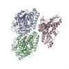
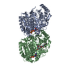
| Title | Refined structure of alpha-beta tubulin from zinc-induced sheets stabilized with taxol | ||||||
 Components Components |
| ||||||
 Keywords Keywords | STRUCTURAL PROTEIN / dimer / GTPase | ||||||
| Function / homology |  Function and homology information Function and homology informationpositive regulation of axon guidance / microtubule-based process / Hydrolases; Acting on acid anhydrides; Acting on GTP to facilitate cellular and subcellular movement / structural constituent of cytoskeleton / microtubule cytoskeleton organization / microtubule cytoskeleton / mitotic cell cycle / nervous system development / microtubule / protein heterodimerization activity ...positive regulation of axon guidance / microtubule-based process / Hydrolases; Acting on acid anhydrides; Acting on GTP to facilitate cellular and subcellular movement / structural constituent of cytoskeleton / microtubule cytoskeleton organization / microtubule cytoskeleton / mitotic cell cycle / nervous system development / microtubule / protein heterodimerization activity / GTPase activity / GTP binding / metal ion binding / cytoplasm / cytosol Similarity search - Function | ||||||
| Biological species |  | ||||||
| Method | ELECTRON CRYSTALLOGRAPHY / electron crystallography /  MOLECULAR REPLACEMENT / cryo EM / Resolution: 3.5 Å MOLECULAR REPLACEMENT / cryo EM / Resolution: 3.5 Å | ||||||
 Authors Authors | Lowe, J. / Li, H. / Downing, K.H. / Nogales, E. | ||||||
 Citation Citation | Journal: J Mol Biol / Year: 2001 Title: Refined structure of alpha beta-tubulin at 3.5 A resolution. Authors: J Löwe / H Li / K H Downing / E Nogales /  Abstract: We present a refined model of the alpha beta-tubulin dimer to 3.5 A resolution. An improved experimental density for the zinc-induced tubulin sheets was obtained by adding 114 electron diffraction ...We present a refined model of the alpha beta-tubulin dimer to 3.5 A resolution. An improved experimental density for the zinc-induced tubulin sheets was obtained by adding 114 electron diffraction patterns at 40-60 degrees tilt and increasing the completeness of structure factor amplitudes to 84.7 %. The refined structure was obtained using maximum-likelihood including phase information from experimental images, and simulated annealing Cartesian refinement to an R-factor of 23.2 and free R-factor of 29.7. The current model includes residues alpha:2-34, alpha:61-439, beta:2-437, one molecule of GTP, one of GDP, and one of taxol, as well as one magnesium ion at the non-exchangeable nucleotide site, and one putative zinc ion near the M-loop in the alpha-tubulin subunit. The acidic C-terminal tails could not be traced accurately, neither could the N-terminal loop including residues 35-60 in the alpha-subunit. There are no major changes in the overall fold of tubulin with respect to the previous structure, testifying to the quality of the initial experimental phases. The overall geometry of the model is, however, greatly improved, and the position of side-chains, especially those of exposed polar/charged groups, is much better defined. Three short protein sequence frame shifts were detected with respect to the non-refined structure. In light of the new model we discuss details of the tubulin structure such as nucleotide and taxol binding sites, lateral contacts in zinc-sheets, and the significance of the location of highly conserved residues. #1: Journal: Nature / Year: 1998Title: Structure of the alpha beta tubulin dimer by electron crystallography Authors: Nogales, E. / Wolf, S.G. / Downing, K.H. | ||||||
| History |
|
- Structure visualization
| Movie |
Movie viewer |
|---|---|
| Structure viewer | Molecule: MolmilJmol/JSmol |
- Downloads & links
-Download
| PDBx/mmCIF format | 1jff.cif.gz | 184.9 KB | Display | PDBx/mmCIF format |
|---|---|---|---|---|
| PDB format | pdb1jff.ent.gz | 139.9 KB | Display | PDB format |
| PDBx/mmJSON format | 1jff.json.gz | Tree view | PDBx/mmJSON format | |
| Others |  Other downloads Other downloads |
-Validation report
| Summary document | 1jff_validation.pdf.gz | 622.3 KB | Display | wwPDB validaton report |
|---|---|---|---|---|
| Full document | 1jff_full_validation.pdf.gz | 724.8 KB | Display | |
| Data in XML | 1jff_validation.xml.gz | 32.6 KB | Display | |
| Data in CIF | 1jff_validation.cif.gz | 46.6 KB | Display | |
| Arichive directory | https://data.pdbj.org/pub/pdb/validation_reports/jf/1jffftp://data.pdbj.org/pub/pdb/validation_reports/jf/1jff | HTTPS FTP |
-Related structure data
| Related structure data |  1tubS S: Starting model for refinement |
|---|---|
| Similar structure data |


-Links
 PDBj
PDBj


- Assembly
| Deposited unit | 
| ||||||||
|---|---|---|---|---|---|---|---|---|---|
| 1 |
| ||||||||
| Unit cell |
|
-Components
-Protein , 2 types, 2 molecules AB
| #1: Protein | Mass: 50107.238 Da / Num. of mol.: 1 / Source method: isolated from a natural source / Source: (natural) |
|---|---|
| #2: Protein | Mass: 49907.770 Da / Num. of mol.: 1 / Source method: isolated from a natural source / Source: (natural) |
-Non-polymers , 5 types, 5 molecules 


| #3: Chemical | ChemComp-ZN /  Mass: 65.409 Da / Num. of mol.: 1 / Source method: obtained synthetically / Formula: Zn Mass: 65.409 Da / Num. of mol.: 1 / Source method: obtained synthetically / Formula: Zn |
|---|---|
| #4: Chemical | ChemComp-MG /  Mass: 24.305 Da / Num. of mol.: 1 / Source method: obtained synthetically / Formula: Mg Mass: 24.305 Da / Num. of mol.: 1 / Source method: obtained synthetically / Formula: Mg |
| #5: Chemical | ChemComp-GTP /  Mass: 523.180 Da / Num. of mol.: 1 / Source method: obtained synthetically / Formula: C10H16N5O14P3 / Comment: GTP, energy-carrying molecule*YM Mass: 523.180 Da / Num. of mol.: 1 / Source method: obtained synthetically / Formula: C10H16N5O14P3 / Comment: GTP, energy-carrying molecule*YM |
| #6: Chemical | ChemComp-GDP /  Type: RNA linking / Mass: 443.201 Da / Num. of mol.: 1 / Source method: obtained synthetically / Formula: C10H15N5O11P2 / Comment: GDP, energy-carrying molecule*YM Type: RNA linking / Mass: 443.201 Da / Num. of mol.: 1 / Source method: obtained synthetically / Formula: C10H15N5O11P2 / Comment: GDP, energy-carrying molecule*YM |
| #7: Chemical | ChemComp-TA1 /  Mass: 853.906 Da / Num. of mol.: 1 / Source method: obtained synthetically / Formula: C47H51NO14 / Comment: medication, chemotherapy*YM Mass: 853.906 Da / Num. of mol.: 1 / Source method: obtained synthetically / Formula: C47H51NO14 / Comment: medication, chemotherapy*YM |
-Details
| Sequence details | THE SAMPLE WAS BOVINE, BUT THE MODELED PROTEIN SEQUENCES ARE FROM SUS SCROFA (PIG). THE AUTHORS ...THE SAMPLE WAS BOVINE, BUT THE MODELED PROTEIN SEQUENCES ARE FROM SUS SCROFA (PIG). THE AUTHORS USED THE SEQUENCES FROM THE MOST ABUNDANT ISOTYPE OF PIG BRAIN TUBULIN. THE RESIDUES IN CHAIN B ARE NOT SEQUENTIAL |
|---|
-Experimental details
-Experiment
| Experiment | Method: ELECTRON CRYSTALLOGRAPHY / Number of used crystals: 200 |
|---|---|
| EM experiment | Aggregation state: 2D ARRAY / 3D reconstruction method: electron crystallography |
- Sample preparation
| Component | Name: Alpha-Beta-tubulin sheets / Type: COMPLEX |
|---|---|
| Specimen | Embedding applied: YES / Shadowing applied: NO / Staining applied: NO / Vitrification applied: YES |
| EM embedding | Material: tannin |
| Vitrification | Cryogen name: NITROGEN |
| Crystal | Description: JEOL 4000 electron microscope was used. Kodak film and gatan CCD were used as detectors. Temperature was 93 Kelvin. |
| Crystal grow | Temperature: 305 K / Method: aberrant polymerization of tubulin / pH: 5.8 Details: Zn++, pH 5.8, aberrant polymerization of tubulin, temperature 305K |
| Crystal grow | *PLUS Method: unknown |
-Data collection
| EM imaging |
| |||||||||||||||
|---|---|---|---|---|---|---|---|---|---|---|---|---|---|---|---|---|
| Image recording | Film or detector model: GENERIC FILM / Details: low dose | |||||||||||||||
| Diffraction |
| |||||||||||||||
| Diffraction source | Source: ELECTRON MICROSCOPE / Type: JEOL 4000 electron microscope / Wavelength: 0.0194 Å | |||||||||||||||
| Detector |
| |||||||||||||||
| Radiation | Protocol: SINGLE WAVELENGTH / Monochromatic (M) / Laue (L): M / Scattering type: electron | |||||||||||||||
| Radiation wavelength | Wavelength: 0.0194 Å / Relative weight: 1 | |||||||||||||||
| Reflection | Resolution: 3.5→20 Å / Num. all: 12422 / Num. obs: 12422 / % possible obs: 73 % / Observed criterion σ(F): 0 / Observed criterion σ(I): 0 / Redundancy: 6 % / Biso Wilson estimate: 40 Å2 / Rmerge(I) obs: 0.25 / Net I/σ(I): 5.4 | |||||||||||||||
| Reflection shell | Resolution: 3.5→3.7 Å / Mean I/σ(I) obs: 2.3 / Num. unique all: 1080 / % possible all: 73 | |||||||||||||||
| Reflection | *PLUS % possible obs: 73 % |
- Processing
| Software |
| ||||||||||||||||||||
|---|---|---|---|---|---|---|---|---|---|---|---|---|---|---|---|---|---|---|---|---|---|
| Refinement | Method to determine structure: MOLECULAR REPLACEMENT Starting model: PDB entry 1TUB Resolution: 3.5→20 Å / Isotropic thermal model: overall anisotropic / σ(F): 0 / σ(I): 0 / Stereochemistry target values: Engh and Huber / Details: constrained
| ||||||||||||||||||||
| Displacement parameters | Biso mean: 41.4 Å2
| ||||||||||||||||||||
| Refinement step | Cycle: LAST / Resolution: 3.5→20 Å
| ||||||||||||||||||||
| Refine LS restraints |
| ||||||||||||||||||||
| LS refinement shell | Resolution: 3.5→3.66 Å
| ||||||||||||||||||||
| Software | *PLUS Name: CNS / Version: 0.9 / Classification: refinement | ||||||||||||||||||||
| Refinement | *PLUS Highest resolution: 3.5 Å / Lowest resolution: 20 Å / σ(F): 0 / Rfactor obs: 0.231 | ||||||||||||||||||||
| Solvent computation | *PLUS | ||||||||||||||||||||
| Displacement parameters | *PLUS Biso mean: 41.4 Å2 | ||||||||||||||||||||
| LS refinement shell | *PLUS Highest resolution: 3.5 Å |
