 Movie
Movie Controller

[English] 日本語
 Yorodumi
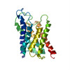
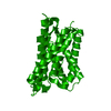
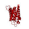
- PDB-1fqy: STRUCTURE OF AQUAPORIN-1 AT 3.8 A RESOLUTION BY ELECTRON CRYSTALL... -
+ Open data

- Basic information
| Entry | Database: PDB / ID: 1fqy | ||||||
|---|---|---|---|---|---|---|---|
| Title | STRUCTURE OF AQUAPORIN-1 AT 3.8 A RESOLUTION BY ELECTRON CRYSTALLOGRAPHY | ||||||
 Components Components | AQUAPORIN-1 | ||||||
 Keywords Keywords | MEMBRANE PROTEIN / WATER CHANNEL / TWO-DIMENSIONAL CRYSTAL | ||||||
| Function / homology |  Function and homology information Function and homology informationnitric oxide transmembrane transporter activity / metanephric descending thin limb development / metanephric proximal straight tubule development / metanephric proximal convoluted tubule segment 2 development / metanephric glomerulus vasculature development / cerebrospinal fluid secretion / lipid digestion / cellular response to salt stress / renal water transport / glycerol transmembrane transporter activity ...nitric oxide transmembrane transporter activity / metanephric descending thin limb development / metanephric proximal straight tubule development / metanephric proximal convoluted tubule segment 2 development / metanephric glomerulus vasculature development / cerebrospinal fluid secretion / lipid digestion / cellular response to salt stress / renal water transport / glycerol transmembrane transporter activity / corticotropin secretion / secretory granule organization / carbon dioxide transmembrane transport / carbon dioxide transmembrane transporter activity / renal water absorption / positive regulation of saliva secretion / Passive transport by Aquaporins / glycerol transmembrane transport / water transmembrane transporter activity / establishment or maintenance of actin cytoskeleton polarity / pancreatic juice secretion / lateral ventricle development / cellular response to mercury ion / potassium ion transmembrane transporter activity / water channel activity / intracellular water homeostasis / cellular response to inorganic substance / intracellularly cGMP-activated cation channel activity / ammonium transmembrane transport / water transport / transepithelial water transport / glomerular filtration / ankyrin-1 complex / ammonium transmembrane transporter activity / camera-type eye morphogenesis / multicellular organismal-level water homeostasis / fibroblast migration / cellular homeostasis / cellular hyperosmotic response / hyperosmotic response / renal water homeostasis / cell volume homeostasis / positive regulation of fibroblast migration / odontogenesis / nitric oxide transport / cGMP-mediated signaling / brush border / potassium channel activity / transmembrane transporter activity / cellular response to nitric oxide / cellular response to retinoic acid / cellular response to cAMP / sensory perception of pain / cellular response to copper ion / ephrin receptor binding / cellular response to dexamethasone stimulus / basal plasma membrane / establishment of localization in cell / brush border membrane / sarcolemma / carbon dioxide transport / wound healing / negative regulation of cysteine-type endopeptidase activity involved in apoptotic process / Erythrocytes take up oxygen and release carbon dioxide / Erythrocytes take up carbon dioxide and release oxygen / potassium ion transport / cellular response to hydrogen peroxide / Vasopressin regulates renal water homeostasis via Aquaporins / cellular response to mechanical stimulus / positive regulation of angiogenesis / cellular response to UV / positive regulation of fibroblast proliferation / apical part of cell / cellular response to hypoxia / basolateral plasma membrane / nuclear membrane / defense response to Gram-negative bacterium / apical plasma membrane / axon / negative regulation of apoptotic process / extracellular exosome / identical protein binding / nucleus / plasma membrane / cytoplasmSimilarity search - Function | ||||||
| Biological species |  Homo sapiens (human) Homo sapiens (human) | ||||||
| Method | ELECTRON CRYSTALLOGRAPHY / electron crystallography / cryo EM / Resolution: 3.8 Å | ||||||
 Authors Authors | Murata, K. / Mitsuoka, K. / Hirai, T. / Walz, T. / Agre, P. / Heymann, J.B. / Engel, A. / Fujiyoshi, Y. | ||||||
 Citation Citation | Journal: Nature / Year: 2000 Title: Structural determinants of water permeation through aquaporin-1. Authors: K Murata / K Mitsuoka / T Hirai / T Walz / P Agre / J B Heymann / A Engel / Y Fujiyoshi /  Abstract: Human red cell AQP1 is the first functionally defined member of the aquaporin family of membrane water channels. Here we describe an atomic model of AQP1 at 3.8A resolution from electron ...Human red cell AQP1 is the first functionally defined member of the aquaporin family of membrane water channels. Here we describe an atomic model of AQP1 at 3.8A resolution from electron crystallographic data. Multiple highly conserved amino-acid residues stabilize the novel fold of AQP1. The aqueous pathway is lined with conserved hydrophobic residues that permit rapid water transport, whereas the water selectivity is due to a constriction of the pore diameter to about 3 A over a span of one residue. The atomic model provides a possible molecular explanation to a longstanding puzzle in physiology-how membranes can be freely permeable to water but impermeable to protons. | ||||||
| History |
|
- Structure visualization
| Movie |
Movie viewer |
|---|---|
| Structure viewer | Molecule: MolmilJmol/JSmol |
- Downloads & links
-Download
| PDBx/mmCIF format | 1fqy.cif.gz | 49.5 KB | Display | PDBx/mmCIF format |
|---|---|---|---|---|
| PDB format | pdb1fqy.ent.gz | 34.4 KB | Display | PDB format |
| PDBx/mmJSON format | 1fqy.json.gz | Tree view | PDBx/mmJSON format | |
| Others |  Other downloads Other downloads |
-Validation report
| Arichive directory | https://data.pdbj.org/pub/pdb/validation_reports/fq/1fqyftp://data.pdbj.org/pub/pdb/validation_reports/fq/1fqy | HTTPS FTP |
|---|
-Related structure data
| Related structure data | |
|---|---|
| Similar structure data |


-Links
 PDBj
PDBj

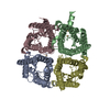
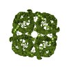
- Assembly
| Deposited unit | 
| ||||||||
|---|---|---|---|---|---|---|---|---|---|
| 1 | 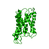
| ||||||||
| Unit cell |
| ||||||||
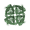
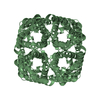
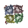
| Details | The biological assembly is a tetramer constructed from chain A |
-Components
| #1: Protein | / AQP1 Mass: 28549.914 Da / Num. of mol.: 1 / Source method: isolated from a natural source / Source: (natural) Homo sapiens (human) / Cell: ERYTHROCYTE / References: UniProt: P29972 |
|---|
-Experimental details
-Experiment
| Experiment | Method: ELECTRON CRYSTALLOGRAPHY / Number of used crystals: 135 |
|---|---|
| EM experiment | Aggregation state: 2D ARRAY / 3D reconstruction method: electron crystallography |
- Sample preparation
| Component | Name: aquaporin / Type: COMPLEX |
|---|---|
| Specimen | Embedding applied: YES / Shadowing applied: NO / Staining applied: NO / Vitrification applied: YES |
| Specimen support | Grid material: MOLYBDENUM |
| EM embedding | Details: 3% / Material: trehalose |
| Crystal grow | Temperature: 298 K / Method: dialysis with continuous flow dialysis machine / pH: 6 Details: Escherichia coli lipids, magnesium chloride, sodium chloride, MES, pH 6.0, Dialysis with continuous flow dialysis machine, temperature 298K |
| Crystal grow | *PLUS Details: Eletron Diffraction |
-Data collection
| Microscopy | Model: JEOL 3000SFF / Details: diffraction patterns and images collected |
|---|---|
| Electron gun | Electron source: FIELD EMISSION GUN / Accelerating voltage: 300 kV / Illumination mode: FLOOD BEAM |
| Electron lens | Mode: DIFFRACTION |
| Specimen holder | Cryogen: HELIUM |
| Image recording | Electron dose: 20 e/Å2 / Film or detector model: GENERIC FILM / Details: digitized using LeafScan45 scanner (Scitex) |
| Image scans | Scanner model: OTHER |
| Diffraction | Mean temperature: 4 K |
| Diffraction source | Source: ELECTRON MICROSCOPE / Type: JEOL 3000 SFF / Wavelength: 0.0197 |
| Detector | Type: GATAN Model 795 MegaScan / Detector: CCD / Date: Jun 7, 1996 |
| Radiation | Protocol: SINGLE WAVELENGTH / Monochromatic (M) / Laue (L): M / Scattering type: electron |
| Radiation wavelength | Wavelength: 0.0197 Å / Relative weight: 1 |
| Reflection | Resolution: 3.8→96 Å / Num. all: 85254 / Num. obs: 85254 / % possible obs: 88 % / Observed criterion σ(F): 0 / Observed criterion σ(I): 0 / Redundancy: 18.8 % / Biso Wilson estimate: 8.14 Å2 / Rmerge(I) obs: 0.495 / Net I/σ(I): 1.1 |
| Reflection shell | Resolution: 3.8→3.9 Å / Redundancy: 13.2 % / Rmerge(I) obs: 0.703 / Num. unique all: 4010 / % possible all: 86.9 |
- Processing
| Software |
| |||||||||||||||||||||||||
|---|---|---|---|---|---|---|---|---|---|---|---|---|---|---|---|---|---|---|---|---|---|---|---|---|---|---|
| 3D reconstruction | Resolution: 3.8 Å / Resolution method: DIFFRACTION PATTERN/LAYERLINES | |||||||||||||||||||||||||
| Refinement | Resolution: 3.8→6 Å / σ(F): 1 / σ(I): 0 / Stereochemistry target values: Engh & Huber Details: For refinement with phase restraint, we used the structure factors merged from both electron diffraction patterns and images. Thus in this file we included the structure factors with phases ...Details: For refinement with phase restraint, we used the structure factors merged from both electron diffraction patterns and images. Thus in this file we included the structure factors with phases used in the refinement. The observed phases were labelled as calculated phases here.
| |||||||||||||||||||||||||
| Refine analyze | Luzzati coordinate error obs: 0.85 Å | |||||||||||||||||||||||||
| Refinement step | Cycle: LAST / Resolution: 3.8→6 Å
| |||||||||||||||||||||||||
| Refine LS restraints |
| |||||||||||||||||||||||||
| LS refinement shell | Resolution: 3.8→3.93 Å / Total num. of bins used: 8
|
