 Movie
Movie Controller


+ Open data

- Basic information
| Entry | Database: EMDB / ID: EMD-1996 | |||||||||
|---|---|---|---|---|---|---|---|---|---|---|
| Title | Bubblegrams reveal the inner body of bacteriophage phiKZ | |||||||||
 Map data Map data | This is the inner body structure of bacteriophage phiKZ. | |||||||||
 Sample Sample |
| |||||||||
 Keywords Keywords | inner body / core protein / asymmetric reconstruction / phiKZ / protein mapping | |||||||||
| Biological species |  Pseudomonas phage phiKZ (virus) Pseudomonas phage phiKZ (virus) | |||||||||
| Method | single particle reconstruction / cryo EM / Resolution: 37.0 Å | |||||||||
 Authors Authors | Wu W / Thomas JA / Cheng N / Black LW / Steven AC | |||||||||
 Citation Citation | Journal: Science / Year: 2012 Title: Bubblegrams reveal the inner body of bacteriophage φKZ. Authors: Weimin Wu / Julie A Thomas / Naiqian Cheng / Lindsay W Black / Alasdair C Steven /  Abstract: Dense packing of macromolecules in cellular compartments and higher-order assemblies makes it difficult to pick out even quite large components in electron micrographs, despite nominally high ...Dense packing of macromolecules in cellular compartments and higher-order assemblies makes it difficult to pick out even quite large components in electron micrographs, despite nominally high resolution. Immunogold labeling and histochemical procedures offer ways to map certain components but are limited in their applicability. Here, we present a differential mapping procedure, based on the physical principle of protein's greater sensitivity to radiation damage compared with that of nucleic acid. | |||||||||
| History |
|
- Structure visualization
| Movie |
 Movie viewer Movie viewer |
|---|---|
| Structure viewer | EM map: SurfViewMolmilJmol/JSmol |
| Supplemental images |

- Downloads & links
-EMDB archive
| Map data | emd_1996.map.gz | 394.8 KB | EMDB map data format | |
|---|---|---|---|---|
| Header (meta data) | emd-1996-v30.xmlemd-1996.xml | 8.7 KB 8.7 KB | Display Display | EMDB header |
| Images | 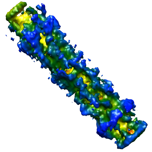 1996.png 1996.png | 141.8 KB | ||
| Archive directory |  http://ftp.pdbj.org/pub/emdb/structures/EMD-1996ftp://ftp.pdbj.org/pub/emdb/structures/EMD-1996 http://ftp.pdbj.org/pub/emdb/structures/EMD-1996ftp://ftp.pdbj.org/pub/emdb/structures/EMD-1996 | HTTPS FTP |
-Validation report
| Summary document | emd_1996_validation.pdf.gz | 191.7 KB | Display | EMDB validaton report |
|---|---|---|---|---|
| Full document | emd_1996_full_validation.pdf.gz | 190.9 KB | Display | |
| Data in XML | emd_1996_validation.xml.gz | 6.4 KB | Display | |
| Arichive directory | https://ftp.pdbj.org/pub/emdb/validation_reports/EMD-1996ftp://ftp.pdbj.org/pub/emdb/validation_reports/EMD-1996 | HTTPS FTP |
-Related structure data
| Similar structure data |
|---|

-Links
| EMDB pages | EMDB (EBI/PDBe) / EMDataResource |
|---|
-Map
| File | Download / File: emd_1996.map.gz / Format: CCP4 / Size: 15.3 MB / Type: IMAGE STORED AS FLOATING POINT NUMBER (4 BYTES) | ||||||||||||||||||||||||||||||||||||||||||||||||||||||||||||||||||||
|---|---|---|---|---|---|---|---|---|---|---|---|---|---|---|---|---|---|---|---|---|---|---|---|---|---|---|---|---|---|---|---|---|---|---|---|---|---|---|---|---|---|---|---|---|---|---|---|---|---|---|---|---|---|---|---|---|---|---|---|---|---|---|---|---|---|---|---|---|---|
| Annotation | This is the inner body structure of bacteriophage phiKZ. | ||||||||||||||||||||||||||||||||||||||||||||||||||||||||||||||||||||
| Voxel size | X=Y=Z: 10.026 Å | ||||||||||||||||||||||||||||||||||||||||||||||||||||||||||||||||||||
| Density |
| ||||||||||||||||||||||||||||||||||||||||||||||||||||||||||||||||||||
| Symmetry | Space group: 1 | ||||||||||||||||||||||||||||||||||||||||||||||||||||||||||||||||||||
| Details | EMDB XML:
CCP4 map header:
| ||||||||||||||||||||||||||||||||||||||||||||||||||||||||||||||||||||
-Supplemental data
- Sample components
-Entire : phiKZ mature phage
| Entire | Name: phiKZ mature phage |
|---|---|
| Components |
|
-Supramolecule #1000: phiKZ mature phage
| Supramolecule | Name: phiKZ mature phage / type: sample / ID: 1000 Oligomeric state: Inner body inside the capsid surrounding by DNA Number unique components: 5 |
|---|
-Supramolecule #1: Pseudomonas phage phiKZ
| Supramolecule | Name: Pseudomonas phage phiKZ / type: virus / ID: 1 / Name.synonym: phiKZ / NCBI-ID: 169683 / Sci species name: Pseudomonas phage phiKZ / Virus type: VIRION / Virus isolate: STRAIN / Virus enveloped: No / Virus empty: No / Syn species name: phiKZ |
|---|---|
| Host (natural) | Organism:   Pseudomonas aeruginosa (bacteria) / synonym: BACTERIA(EUBACTERIA) Pseudomonas aeruginosa (bacteria) / synonym: BACTERIA(EUBACTERIA) |
-Experimental details
-Structure determination
| Method | cryo EM |
|---|---|
 Processing Processing | single particle reconstruction |
| Aggregation state | particle |
-Sample preparation
| Vitrification | Cryogen name: ETHANE / Chamber humidity: 40 % / Chamber temperature: 100 K / Instrument: OTHER |
|---|
- Electron microscopy
| Microscope | FEI/PHILIPS CM200FEG |
|---|---|
| Temperature | Average: 100 K |
| Alignment procedure | Legacy - Astigmatism: condenser and objective lens astigmatism |
| Details | Image pairs. First low dose image, 12 electrons per angstrom squared, then high dose to get radiation damage (same dose but longer time), dose about 5-fold of low dose. |
| Date | Jan 20, 2011 |
| Image recording | Category: FILM / Film or detector model: KODAK SO-163 FILM / Digitization - Scanner: NIKON SUPER COOLSCAN 9000 / Digitization - Sampling interval: 6.35 µm / Number real images: 95 / Average electron dose: 12 e/Å2 / Bits/pixel: 16 |
| Electron beam | Acceleration voltage: 120 kV / Electron source:  FIELD EMISSION GUN FIELD EMISSION GUN |
| Electron optics | Calibrated magnification: 38000 / Illumination mode: SPOT SCAN / Imaging mode: OTHER / Cs: 2.0 mm / Nominal defocus max: 1.5 µm / Nominal defocus min: 0.8 µm / Nominal magnification: 38000 |
| Sample stage | Specimen holder: Eucentric / Specimen holder model: GATAN LIQUID NITROGEN |
-Image processing
| CTF correction | Details: Micrograph |
|---|---|
| Final reconstruction | Applied symmetry - Point group: C1 (asymmetric) / Algorithm: OTHER / Resolution.type: BY AUTHOR / Resolution: 37.0 Å / Resolution method: FSC 0.5 CUT-OFF / Software - Name: EMAN Details: Projection match method was used to determine the unique vertex and the orientation of tail. The inner body was solved directly from 2D data without using any initial model. Number images used: 2775 |
