 Movie
Movie Controller

[English] 日本語
 Yorodumi
- EMDB-1924: Ribosome Assembly Factors Prevent Premature Translation Initiatio... -
+ Open data

- Basic information
| Entry | Database: EMDB / ID: EMD-1924 | |||||||||
|---|---|---|---|---|---|---|---|---|---|---|
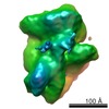
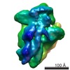
| Title | Ribosome Assembly Factors Prevent Premature Translation Initiation by 40S Assembly Intermediates | |||||||||
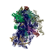
 Map data Map data | Surface rendered Ltv1 deletion map | |||||||||
 Sample Sample |
| |||||||||
 Keywords Keywords | pre-40S / 40S intermediate / Ltv1 deletion | |||||||||
| Biological species |  | |||||||||
| Method | single particle reconstruction / cryo EM / Resolution: 20.0 Å | |||||||||
 Authors Authors | Strunk BS / Loucks CR / Su M / Vashisth H / Cheng S / Schilling J / BrooksIII CL / Karbstein K / Skiniotis G | |||||||||
 Citation Citation | Journal: Science / Year: 2011 Title: Ribosome assembly factors prevent premature translation initiation by 40S assembly intermediates. Authors: Bethany S Strunk / Cherisse R Loucks / Min Su / Harish Vashisth / Shanshan Cheng / Justin Schilling / Charles L Brooks / Katrin Karbstein / Georgios Skiniotis /  Abstract: Ribosome assembly in eukaryotes requires approximately 200 essential assembly factors (AFs) and occurs through ordered events that initiate in the nucleolus and culminate in the cytoplasm. Here, we ...Ribosome assembly in eukaryotes requires approximately 200 essential assembly factors (AFs) and occurs through ordered events that initiate in the nucleolus and culminate in the cytoplasm. Here, we present the electron cryo-microscopy (cryo-EM) structure of a late cytoplasmic 40S ribosome assembly intermediate from Saccharomyces cerevisiae at 18 angstrom resolution. We obtained cryo-EM reconstructions of preribosomal complexes lacking individual components to define the positions of all seven AFs bound to this intermediate. These late-binding AFs are positioned to prevent each step in the translation initiation pathway. Together, they obstruct the binding sites for initiation factors, prevent the opening of the messenger RNA channel, block 60S subunit joining, and disrupt the decoding site. These redundant mechanisms probably ensure that pre-40S particles do not enter the translation pathway, which would result in their rapid degradation. | |||||||||
| History |
|
- Structure visualization
| Movie |
 Movie viewer Movie viewer |
|---|---|
| Structure viewer | EM map: SurfViewMolmilJmol/JSmol |
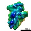
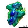
| Supplemental images |

- Downloads & links
-EMDB archive
| Map data | emd_1924.map.gz | 11.4 MB | EMDB map data format | |
|---|---|---|---|---|
| Header (meta data) | emd-1924-v30.xmlemd-1924.xml | 10 KB 10 KB | Display Display | EMDB header |
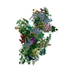
| Images | 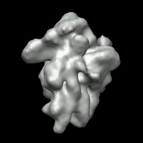 1924.png 1924.png | 91.5 KB | ||
| Archive directory |  http://ftp.pdbj.org/pub/emdb/structures/EMD-1924ftp://ftp.pdbj.org/pub/emdb/structures/EMD-1924 http://ftp.pdbj.org/pub/emdb/structures/EMD-1924ftp://ftp.pdbj.org/pub/emdb/structures/EMD-1924 | HTTPS FTP |
-Validation report
| Summary document | emd_1924_validation.pdf.gz | 207.1 KB | Display | EMDB validaton report |
|---|---|---|---|---|
| Full document | emd_1924_full_validation.pdf.gz | 206.3 KB | Display | |
| Data in XML | emd_1924_validation.xml.gz | 5.9 KB | Display | |
| Arichive directory | https://ftp.pdbj.org/pub/emdb/validation_reports/EMD-1924ftp://ftp.pdbj.org/pub/emdb/validation_reports/EMD-1924 | HTTPS FTP |
-Related structure data
-Links
| EMDB pages | EMDB (EBI/PDBe) / EMDataResource |
|---|---|
| Related items in Molecule of the Month |

-Map
| File | Download / File: emd_1924.map.gz / Format: CCP4 / Size: 15.3 MB / Type: IMAGE STORED AS FLOATING POINT NUMBER (4 BYTES) | ||||||||||||||||||||||||||||||||||||||||||||||||||||||||||||||||||||
|---|---|---|---|---|---|---|---|---|---|---|---|---|---|---|---|---|---|---|---|---|---|---|---|---|---|---|---|---|---|---|---|---|---|---|---|---|---|---|---|---|---|---|---|---|---|---|---|---|---|---|---|---|---|---|---|---|---|---|---|---|---|---|---|---|---|---|---|---|---|
| Annotation | Surface rendered Ltv1 deletion map | ||||||||||||||||||||||||||||||||||||||||||||||||||||||||||||||||||||
| Voxel size | X=Y=Z: 2.24 Å | ||||||||||||||||||||||||||||||||||||||||||||||||||||||||||||||||||||
| Density |
| ||||||||||||||||||||||||||||||||||||||||||||||||||||||||||||||||||||
| Symmetry | Space group: 1 | ||||||||||||||||||||||||||||||||||||||||||||||||||||||||||||||||||||
| Details | EMDB XML:
CCP4 map header:
| ||||||||||||||||||||||||||||||||||||||||||||||||||||||||||||||||||||
-Supplemental data
- Sample components
-Entire : S. cerevisiae pre-40S ribosomal particle with Ltv1 deletion
| Entire | Name: S. cerevisiae pre-40S ribosomal particle with Ltv1 deletion |
|---|---|
| Components |
|
-Supramolecule #1000: S. cerevisiae pre-40S ribosomal particle with Ltv1 deletion
| Supramolecule | Name: S. cerevisiae pre-40S ribosomal particle with Ltv1 deletion type: sample / ID: 1000 / Details: Monodisperse / Number unique components: 1 |
|---|---|
| Molecular weight | Theoretical: 1.4 MDa |
-Supramolecule #1: pre-40S
| Supramolecule | Name: pre-40S / type: complex / ID: 1 / Name.synonym: pre-40S / Recombinant expression: No / Ribosome-details: ribosome-eukaryote: SSU 40S |
|---|---|
| Source (natural) | Organism: |
| Molecular weight | Theoretical: 1.4 MDa |
-Experimental details
-Structure determination
| Method | cryo EM |
|---|---|
 Processing Processing | single particle reconstruction |
| Aggregation state | particle |
-Sample preparation
| Concentration | 1.5 mg/mL |
|---|---|
| Buffer | pH: 7.5 Details: 50mM Tris-Cl, 100mM NaCl, 10mM MgCl2, 0.075% NP40, 1mM imidazole, 2mM EGTA, 10 mM BME |
| Grid | Details: Quantifoil |
| Vitrification | Cryogen name: ETHANE / Chamber humidity: 100 % / Chamber temperature: 85 K / Instrument: OTHER / Details: Vitrification instrument: Vitrobot / Method: Blot for 2 seconds before plunging |
- Electron microscopy
| Microscope | FEI TECNAI F20 |
|---|---|
| Temperature | Min: 89 K / Max: 89 K / Average: 89 K |
| Alignment procedure | Legacy - Astigmatism: Objective lens astigmatism was corrected at 135,000 times magnification |
| Image recording | Category: CCD / Film or detector model: GATAN ULTRASCAN 4000 (4k x 4k) / Digitization - Sampling interval: 2.24 µm / Number real images: 350 / Average electron dose: 16 e/Å2 |
| Electron beam | Acceleration voltage: 200 kV / Electron source:  FIELD EMISSION GUN FIELD EMISSION GUN |
| Electron optics | Calibrated magnification: 66964 / Illumination mode: FLOOD BEAM / Imaging mode: BRIGHT FIELD / Cs: 2 mm / Nominal defocus max: 4.0 µm / Nominal defocus min: 1.5 µm |
| Sample stage | Specimen holder: Side entry liquid nitrogen-cooled cryo specimen holder Specimen holder model: OTHER |
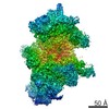
| Experimental equipment |  Model: Tecnai F20 / Image courtesy: FEI Company |
-Image processing
| Details | Manual particle selection |
|---|---|
| CTF correction | Details: Each micrograph |
| Final reconstruction | Applied symmetry - Point group: C1 (asymmetric) / Algorithm: OTHER / Resolution.type: BY AUTHOR / Resolution: 20.0 Å / Resolution method: FSC 0.5 CUT-OFF / Software - Name: EMAN / Details: Final map was filtered to 20A resolution / Number images used: 10235 |
| Final angle assignment | Details: EMAN convention |
-Atomic model buiding 1
| Initial model | PDB ID:  3o2z |
|---|---|
| Software | Name: NAMD |
| Details | Protocol: MDFF |
| Refinement | Space: REAL / Protocol: FLEXIBLE FIT / Target criteria: CC |
