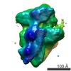
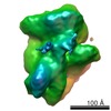
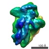
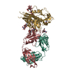
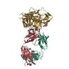
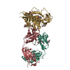
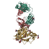
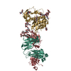
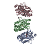
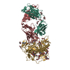
Journal: Science / Year: 2011 Title: Ribosome assembly factors prevent premature translation initiation by 40S assembly intermediates. Authors: Bethany S Strunk / Cherisse R Loucks / Min Su / Harish Vashisth / Shanshan Cheng / Justin Schilling / Charles L Brooks / Katrin Karbstein / Georgios Skiniotis / Abstract: Ribosome assembly in eukaryotes requires approximately 200 essential assembly factors (AFs) and occurs through ordered events that initiate in the nucleolus and culminate in the cytoplasm. Here, we ...Ribosome assembly in eukaryotes requires approximately 200 essential assembly factors (AFs) and occurs through ordered events that initiate in the nucleolus and culminate in the cytoplasm. Here, we present the electron cryo-microscopy (cryo-EM) structure of a late cytoplasmic 40S ribosome assembly intermediate from Saccharomyces cerevisiae at 18 angstrom resolution. We obtained cryo-EM reconstructions of preribosomal complexes lacking individual components to define the positions of all seven AFs bound to this intermediate. These late-binding AFs are positioned to prevent each step in the translation initiation pathway. Together, they obstruct the binding sites for initiation factors, prevent the opening of the messenger RNA channel, block 60S subunit joining, and disrupt the decoding site. These redundant mechanisms probably ensure that pre-40S particles do not enter the translation pathway, which would result in their rapid degradation.
History
Deposition
Jul 4, 2011
-
Header (metadata) release
Nov 4, 2011
-
Map release
Nov 4, 2011
-
Update
Oct 1, 2014
-
Current status
Oct 1, 2014
Processing site: PDBe / Status: Released
-
Structure visualization
Movie
Surface view with section colored by density value
In the structure databanks used in Yorodumi, some data are registered as the other names, "COVID-19 virus" and "2019-nCoV". Here are the details of the virus and the list of structure data.
Jan 31, 2019. EMDB accession codes are about to change! (news from PDBe EMDB page)
EMDB accession codes are about to change! (news from PDBe EMDB page)
The allocation of 4 digits for EMDB accession codes will soon come to an end. Whilst these codes will remain in use, new EMDB accession codes will include an additional digit and will expand incrementally as the available range of codes is exhausted. The current 4-digit format prefixed with “EMD-” (i.e. EMD-XXXX) will advance to a 5-digit format (i.e. EMD-XXXXX), and so on. It is currently estimated that the 4-digit codes will be depleted around Spring 2019, at which point the 5-digit format will come into force.
The EM Navigator/Yorodumi systems omit the EMD- prefix.
Related info.:Q: What is EMD? / ID/Accession-code notation in Yorodumi/EM Navigator
Yorodumi is a browser for structure data from EMDB, PDB, SASBDB, etc.
This page is also the successor to EM Navigator detail page, and also detail information page/front-end page for Omokage search.
The word "yorodu" (or yorozu) is an old Japanese word meaning "ten thousand". "mi" (miru) is to see.
Related info.:EMDB / PDB / SASBDB / Comparison of 3 databanks / Yorodumi Search / Aug 31, 2016. New EM Navigator & Yorodumi / Yorodumi Papers / Jmol/JSmol / Function and homology information / Changes in new EM Navigator and Yorodumi
 Movie
Movie Controller

 Yorodumi
 Open data
 Basic information
 Map data
 Sample
 Keywords
 Function and homology information
 Authors
 Citation
 Structure visualization
 UCSF Chimera
 Downloads & links
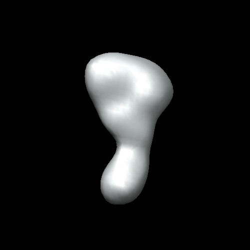 EMD-1922.png
 http://ftp.pdbj.org/pub/emdb/structures/EMD-1922
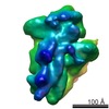


 Sample components
 Processing
 Electron microscopy