Movie
Movie Controller

[English] 日本語
 Yorodumi
- EMDB-1895: Cryo-electron Microscopy Structure of the 30S Subunit in Complex ... -
+ Open data

- Basic information
| Entry | Database: EMDB / ID: EMD-1895 | |||||||||
|---|---|---|---|---|---|---|---|---|---|---|
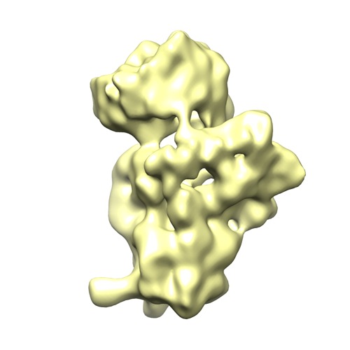
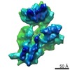
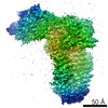
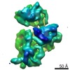
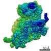
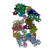
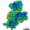
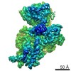
| Title | Cryo-electron Microscopy Structure of the 30S Subunit in Complex with the YjeQ Biogenesis Factor | |||||||||
 Map data Map data | YjeQ in complex with the 30S ribosomal subunit | |||||||||
 Sample Sample |
| |||||||||
 Keywords Keywords |  Ribosome assembly / 30S subunit / YjeQ protein / RsgA protein / GTPase Ribosome assembly / 30S subunit / YjeQ protein / RsgA protein / GTPase | |||||||||
| Function / homology |  Function and homology information Function and homology informationmRNA base-pairing translational repressor activity / ornithine decarboxylase inhibitor activity / Hydrolases; Acting on acid anhydrides; In phosphorus-containing anhydrides / misfolded RNA binding / transcription antitermination factor activity, RNA binding / Group I intron splicing / RNA folding / four-way junction DNA binding / negative regulation of translational initiation / regulation of mRNA stability ...mRNA base-pairing translational repressor activity / ornithine decarboxylase inhibitor activity / Hydrolases; Acting on acid anhydrides; In phosphorus-containing anhydrides / misfolded RNA binding / transcription antitermination factor activity, RNA binding / Group I intron splicing / RNA folding / four-way junction DNA binding / negative regulation of translational initiation / regulation of mRNA stability / mRNA regulatory element binding translation repressor activity / transcription elongation factor complex / positive regulation of RNA splicing / DNA endonuclease activity / regulation of DNA-templated transcription elongation / transcription antitermination / maintenance of translational fidelity / DNA-templated transcription termination / mRNA 5'-UTR binding / ribosomal small subunit biogenesis / small ribosomal subunit rRNA binding / ribosomal small subunit assembly / cytosolic small ribosomal subunit / ribosome biogenesis / regulation of translation / small ribosomal subunit / cytoplasmic translation / negative regulation of translation / tRNA binding / molecular adaptor activity / rRNA binding / ribosome / structural constituent of ribosome / translation / response to antibiotic / mRNA binding / GTPase activity / GTP binding / RNA binding / zinc ion binding / membrane / metal ion binding / cytosol / cytoplasmSimilarity search - Function | |||||||||
| Biological species |  Escherichia coli (E. coli) Escherichia coli (E. coli) | |||||||||
| Method | single particle reconstruction / cryo EM / Resolution: 16.5 Å | |||||||||
 Authors Authors | Jomaa A / Stewart G / Mears JA / Kireeva I / Brown ED / Ortega J | |||||||||
 Citation Citation | Journal: RNA / Year: 2011 Title: Cryo-electron microscopy structure of the 30S subunit in complex with the YjeQ biogenesis factor. Authors: Ahmad Jomaa / Geordie Stewart / Jason A Mears / Inga Kireeva / Eric D Brown / Joaquin Ortega /  Abstract: YjeQ is a protein broadly conserved in bacteria containing an N-terminal oligonucleotide/oligosaccharide fold (OB-fold) domain, a central GTPase domain, and a C-terminal zinc-finger domain. YjeQ ...YjeQ is a protein broadly conserved in bacteria containing an N-terminal oligonucleotide/oligosaccharide fold (OB-fold) domain, a central GTPase domain, and a C-terminal zinc-finger domain. YjeQ binds tightly and stoichiometrically to the 30S subunit, which stimulates its GTPase activity by 160-fold. Despite growing evidence for the involvement of the YjeQ protein in bacterial 30S subunit assembly, the specific function and mechanism of this protein remain unclear. Here, we report the costructure of YjeQ with the 30S subunit obtained by cryo-electron microscopy. The costructure revealed that YjeQ interacts simultaneously with helix 44, the head and the platform of the 30S subunit. This binding location of YjeQ in the 30S subunit suggests a chaperone role in processing of the 3' end of the rRNA as well as in mediating the correct orientation of the main domains of the 30S subunit. In addition, the YjeQ binding site partially overlaps with the interaction site of initiation factors 2 and 3, and upon binding, YjeQ covers three inter-subunit bridges that are important for the association of the 30S and 50S subunits. Hence, our structure suggests that YjeQ may assist in ribosome maturation by preventing premature formation of the translation initiation complex and association with the 50S subunit. Together, these results support a role for YjeQ in the late stages of 30S maturation. | |||||||||
| History |
|
- Structure visualization
| Movie |
Movie viewer |
|---|---|
| Structure viewer | EM map: SurfViewMolmilJmol/JSmol |
| Supplemental images |
- Downloads & links
-EMDB archive
| Map data | emd_1895.map.gz | 7.4 MB | EMDB map data format | |
|---|---|---|---|---|
| Header (meta data) | emd-1895-v30.xmlemd-1895.xml | 11.5 KB 11.5 KB | Display Display | EMDB header |
| Images | YjeQ+30S.tif | 136.9 KB | ||
| Archive directory |  http://ftp.pdbj.org/pub/emdb/structures/EMD-1895ftp://ftp.pdbj.org/pub/emdb/structures/EMD-1895 http://ftp.pdbj.org/pub/emdb/structures/EMD-1895ftp://ftp.pdbj.org/pub/emdb/structures/EMD-1895 | HTTPS FTP |
-Related structure data
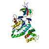
| Related structure data |  4a2iMC M: atomic model generated by this map C: citing same article ( |
|---|---|
| Similar structure data |
-Links
| EMDB pages | EMDB (EBI/PDBe) / EMDataResource |
|---|---|
| Related items in Molecule of the Month |


-Map
| File | Download / File: emd_1895.map.gz / Format: CCP4 / Size: 7.8 MB / Type: IMAGE STORED AS FLOATING POINT NUMBER (4 BYTES) | ||||||||||||||||||||||||||||||||||||||||||||||||||||||||||||||||||||
|---|---|---|---|---|---|---|---|---|---|---|---|---|---|---|---|---|---|---|---|---|---|---|---|---|---|---|---|---|---|---|---|---|---|---|---|---|---|---|---|---|---|---|---|---|---|---|---|---|---|---|---|---|---|---|---|---|---|---|---|---|---|---|---|---|---|---|---|---|---|
| Annotation | YjeQ in complex with the 30S ribosomal subunit | ||||||||||||||||||||||||||||||||||||||||||||||||||||||||||||||||||||
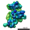
| Voxel size | X=Y=Z: 2.54 Å | ||||||||||||||||||||||||||||||||||||||||||||||||||||||||||||||||||||
| Density |
| ||||||||||||||||||||||||||||||||||||||||||||||||||||||||||||||||||||
| Symmetry | Space group: 1 | ||||||||||||||||||||||||||||||||||||||||||||||||||||||||||||||||||||
| Details | EMDB XML:
CCP4 map header:
| ||||||||||||||||||||||||||||||||||||||||||||||||||||||||||||||||||||
-Supplemental data
- Sample components
-Entire : Escherichia coli 30S ribosomal subunit in complex with the YjeQ p...
| Entire | Name: Escherichia coli 30S ribosomal subunit in complex with the YjeQ protein |
|---|---|
| Components |
|
-Supramolecule #1000: Escherichia coli 30S ribosomal subunit in complex with the YjeQ p...
| Supramolecule | Name: Escherichia coli 30S ribosomal subunit in complex with the YjeQ protein type: sample / ID: 1000 / Details: 30S was mixed with YjeQ in 1-to-5 molar ratio / Number unique components: 2 |
|---|---|
| Molecular weight | Theoretical: 900 KDa |
-Supramolecule #1: 30S ribosomal subunit
| Supramolecule | Name: 30S ribosomal subunit / type: complex / ID: 1 / Name.synonym: 30S subunit / Recombinant expression: No / Ribosome-details: ribosome-prokaryote: SSU 30S |
|---|---|
| Source (natural) | Organism: Escherichia coli (E. coli) |
| Molecular weight | Theoretical: 900 KDa |
-Macromolecule #1: RsgA, YjeQ
| Macromolecule | Name: RsgA, YjeQ / type: protein_or_peptide / ID: 1 / Name.synonym: Assembly factor / Oligomeric state: Monomer / Recombinant expression: Yes |
|---|---|
| Source (natural) | Organism: Escherichia coli (E. coli) / Strain: BW25113 / Cell: Cytoplasm |
| Molecular weight | Experimental: 39 KDa |
| Recombinant expression | Organism: Escherichia coli (E. coli) / Recombinant plasmid: pDEST17-YjeQ |
-Experimental details
-Structure determination
| Method | cryo EM |
|---|---|
 Processing Processing | single particle reconstruction |
| Aggregation state | particle |
-Sample preparation
| Concentration | 0.9 mg/mL |
|---|---|
| Buffer | pH: 7.5 Details: 10 mM Tris-HCl pH 7.5, 10 mM magnesium acetate, 60 mM NH4Cl, 3 mM 2-mercaptoethanol. |
| Grid | Details: 200 mesh copper grid |
| Vitrification | Cryogen name: ETHANE / Chamber humidity: 100 % / Chamber temperature: 100 K / Instrument: FEI VITROBOT MARK III / Details: Vitrification instrument: FEI Vitrobot III / Method: Blot for 7 seconds |
- Electron microscopy
| Microscope | JEOL 2010F |
|---|---|
| Electron beam | Acceleration voltage: 200 kV / Electron source: FIELD EMISSION GUN |
| Electron optics | Calibrated magnification: 50000 / Illumination mode: FLOOD BEAM / Imaging mode: BRIGHT FIELDBright-field microscopy / Cs: 1.0 mm / Nominal defocus max: 3.9 µm / Nominal defocus min: 0.65 µm / Nominal magnification: 50000 |
| Sample stage | Specimen holder: Side entry liquid nitrogen-cooled cryo specimen holder.This holder operates in the temperature range from -175 C to ambient Specimen holder model: GATAN LIQUID NITROGEN |
| Temperature | Average: 100 K |
| Alignment procedure | Legacy - Astigmatism: Corrected at 200000 times magnification |
| Date | May 12, 2010 |
| Image recording | Category: FILM / Film or detector model: KODAK SO-163 FILM / Digitization - Scanner: NIKON SUPER COOLSCAN 9000 / Digitization - Sampling interval: 2.54 µm / Number real images: 150 / Average electron dose: 15 e/Å2 / Bits/pixel: 8 |
-Image processing
| CTF correction | Details: Each micrograph |
|---|---|
| Final reconstruction | Applied symmetry - Point group: C1 (asymmetric) / Algorithm: OTHER / Resolution.type: BY AUTHOR / Resolution: 16.5 Å / Resolution method: FSC 0.5 CUT-OFF / Software - Name: Xmipp / Number images used: 16228 |
| Details | Particles were picked with Boxer. |
-Atomic model buiding 1
| Initial model | PDB ID:  2avy |
|---|---|
| Software | Name: Chimera |
| Details | Protocol: Rigid Body. Each domain of YjeQ was fitted separately |
| Refinement | Space: REAL / Protocol: RIGID BODY FIT / Target criteria: R-factor |
| Output model | PDB-4a2i: |
-Atomic model buiding 2
| Initial model | PDB ID: |
|---|---|
| Software | Name: Chimera |
| Details | Protocol: Rigid Body. Each domain of YjeQ was fitted separately |
| Refinement | Space: REAL / Protocol: RIGID BODY FIT / Target criteria: R-factor |
| Output model | PDB-4a2i: |