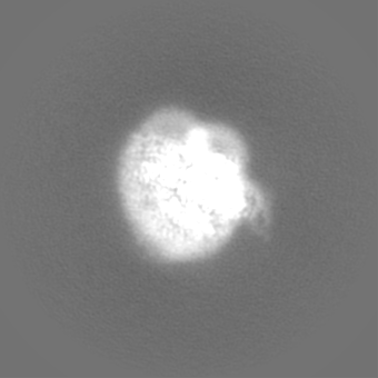
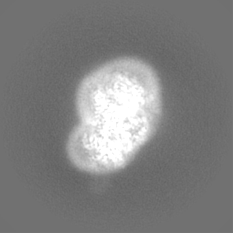
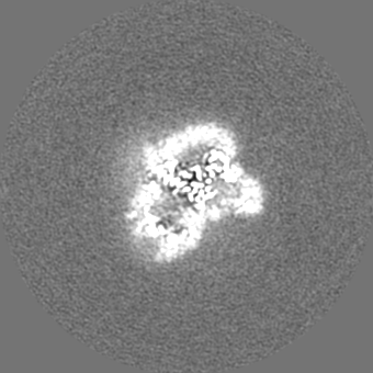
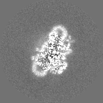
- EMDB-18848: Cryo-EM Structure of native Photosystem II assembly intermediate ... -
+
Open data
ID or keywords:
Loading...
-
Basic information
Entry
Database: EMDB / ID: EMD-18848
Title
Cryo-EM Structure of native Photosystem II assembly intermediate fromChlamydomonas reinhardtii
Map data
Sample
Complex: Photosystem II assembly intermediate from Chlamydomonas reinhardtii
Protein or peptide: x 11 types
Ligand: x 13 types
Keywords
Photosystem II / photosynthesis / biogenesis of PSII / assembly intermediate / CryoEM / assembly factor / Chlamydomonas reinhardtii
Function / homology
Function and homology information
photosystem II repair / chloroplast thylakoid lumen / photosystem II assembly / oxygen evolving activity / photosystem II / photosystem II reaction center / oxidoreductase activity, acting on diphenols and related substances as donors, oxygen as acceptor / photosynthetic electron transport chain / response to herbicide / photosystem II ...photosystem II repair / chloroplast thylakoid lumen / photosystem II assembly / oxygen evolving activity / photosystem II / photosystem II reaction center / oxidoreductase activity, acting on diphenols and related substances as donors, oxygen as acceptor / photosynthetic electron transport chain / response to herbicide / photosystem II / photosynthetic electron transport in photosystem II / chlorophyll binding / chloroplast thylakoid membrane / electron transporter, transferring electrons within the cyclic electron transport pathway of photosynthesis activity / phosphate ion binding / photosynthesis / electron transfer activity / protein stabilization / iron ion binding / heme binding / metal ion binding Similarity search - Function
Photosystem II Pbs27 / Photosystem II Pbs27 superfamily / Photosystem II Pbs27 / Photosystem II reaction centre protein Ycf12 / Photosystem II complex subunit Ycf12 / Photosystem II CP43 reaction centre protein superfamily / Photosystem II PsbK / Photosystem II CP43 reaction centre protein / Photosystem II PsbK superfamily / Photosystem II 4 kDa reaction centre component ...Photosystem II Pbs27 / Photosystem II Pbs27 superfamily / Photosystem II Pbs27 / Photosystem II reaction centre protein Ycf12 / Photosystem II complex subunit Ycf12 / Photosystem II CP43 reaction centre protein superfamily / Photosystem II PsbK / Photosystem II CP43 reaction centre protein / Photosystem II PsbK superfamily / Photosystem II 4 kDa reaction centre component / Photosystem II CP47 reaction centre protein / Photosystem II PsbI / Photosystem II PsbI superfamily / Photosystem II reaction centre I protein (PSII 4.8 kDa protein) / Photosystem II reaction centre protein H / Photosystem II protein D1 / Photosystem II D2 protein / Photosystem II cytochrome b559, conserved site / Photosystem II cytochrome b559, alpha subunit / Photosystem II cytochrome b559, beta subunit / Photosystem II cytochrome b559, N-terminal / Photosystem II cytochrome b559, alpha subunit, lumenal region / Photosystem II reaction centre protein H superfamily / Photosystem II cytochrome b559, alpha subunit superfamily / Cytochrome b559, alpha (gene psbE) and beta (gene psbF)subunits / Lumenal portion of Cytochrome b559, alpha (gene psbE) subunit / Photosystem II 10 kDa phosphoprotein / Cytochrome b559 subunits heme-binding site signature. / Photosystem antenna protein-like / Photosystem antenna protein-like superfamily / Photosystem II protein / Photosynthetic reaction centre, L/M / Photosystem II protein D1/D2 superfamily / Photosynthetic reaction centre protein / Photosynthetic reaction center proteins signature. Similarity search - Domain/homology
Uncharacterized protein / Photosystem II D2 protein / Photosystem II protein D1 / Photosystem II CP43 reaction center protein / Photosystem II reaction center protein K / Photosystem II reaction center protein H / Photosystem II CP47 reaction center protein / Cytochrome b559 subunit alpha / Photosystem II reaction center protein Psb30 / Photosystem II reaction center protein I / Cytochrome b559 subunit beta Similarity search - Component
Biological species
Chlamydomonas reinhardtii (plant)
Method
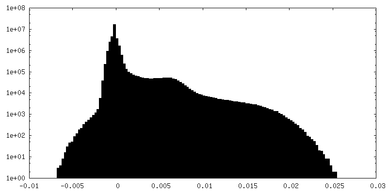
single particle reconstruction / cryo EM / Resolution: 2.9 Å
Journal: Front Plant Sci / Year: 2023 Title: Structure of native photosystem II assembly intermediate from . Authors: Mariia Fadeeva / Daniel Klaiman / Eaazhisai Kandiah / Nathan Nelson / Abstract: Photosystem II (PSII) is a dimer consisting of at least 13 nuclear-encoded and four chloroplast-encoded protein subunits that collectively function as a sunlight-driven oxidoreductase. In this ... Photosystem II (PSII) is a dimer consisting of at least 13 nuclear-encoded and four chloroplast-encoded protein subunits that collectively function as a sunlight-driven oxidoreductase. In this study, we present the inaugural structure of a green alga PSII assembly intermediate (pre-PSII-int). This intermediate was isolated from chloroplast membranes of the temperature-sensitive mutant TSP4, cultivated for 14 hours at a non-permissive temperature. The assembly state comprises a monomer containing subunits A, B, C, D, E, F, H, I, K, and two novel assembly factors, Psb1 and Psb2. Psb1 is identified as a novel transmembrane helix located adjacent to PsbE and PsbF (cytochrome b559). The absence of PsbJ, typically found in mature PSII close to this position, indicates that Psb1 functions as an assembly factor. Psb2 is an eukaryotic homolog of the cyanobacterial assembly factor Psb27. The presence of iron, coupled with the absence of Q, Q, and the manganese cluster, implies a protective mechanism against photodamage and provides insights into the intricate assembly process.
Film or detector model: GATAN K3 BIOQUANTUM (6k x 4k) / Number grids imaged: 3 / Number real images: 25758 / Average exposure time: 1.8 sec. / Average electron dose: 1.07 e/Å2
In the structure databanks used in Yorodumi, some data are registered as the other names, "COVID-19 virus" and "2019-nCoV". Here are the details of the virus and the list of structure data.
Jan 31, 2019. EMDB accession codes are about to change! (news from PDBe EMDB page)
EMDB accession codes are about to change! (news from PDBe EMDB page)
The allocation of 4 digits for EMDB accession codes will soon come to an end. Whilst these codes will remain in use, new EMDB accession codes will include an additional digit and will expand incrementally as the available range of codes is exhausted. The current 4-digit format prefixed with “EMD-” (i.e. EMD-XXXX) will advance to a 5-digit format (i.e. EMD-XXXXX), and so on. It is currently estimated that the 4-digit codes will be depleted around Spring 2019, at which point the 5-digit format will come into force.
The EM Navigator/Yorodumi systems omit the EMD- prefix.
Related info.:Q: What is EMD? / ID/Accession-code notation in Yorodumi/EM Navigator
Yorodumi is a browser for structure data from EMDB, PDB, SASBDB, etc.
This page is also the successor to EM Navigator detail page, and also detail information page/front-end page for Omokage search.
The word "yorodu" (or yorozu) is an old Japanese word meaning "ten thousand". "mi" (miru) is to see.
Related info.:EMDB / PDB / SASBDB / Comparison of 3 databanks / Yorodumi Search / Aug 31, 2016. New EM Navigator & Yorodumi / Yorodumi Papers / Jmol/JSmol / Function and homology information / Changes in new EM Navigator and Yorodumi
 Movie
Movie Controller

 Yorodumi
 Open data
 Basic information

 Map data
 Sample
 Keywords
 Function and homology information

 Authors
 Israel, 1 items
  Citation
 Structure visualization
 Downloads & links
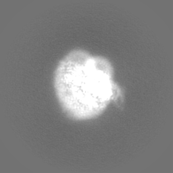
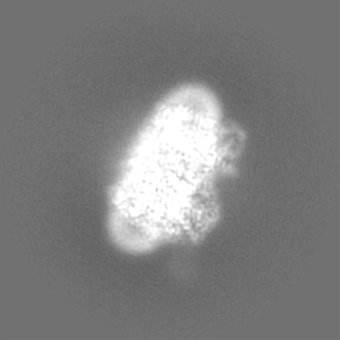
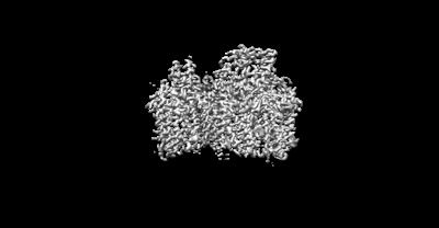 emd_18848.png
 http://ftp.pdbj.org/pub/emdb/structures/EMD-18848


 Z
Z Y
Y X
X


 Sample components


 Processing
 Electron microscopy


