 Movie
Movie Controller

[English] 日本語
 Yorodumi
- EMDB-1882: Mutually-induced conformational switching of RNA and coat protein... -
+ Open data

- Basic information
| Entry | Database: EMDB / ID: EMD-1882 | |||||||||
|---|---|---|---|---|---|---|---|---|---|---|
| Title | Mutually-induced conformational switching of RNA and coat protein underpins efficient assembly of a viral capsid. | |||||||||
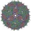
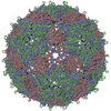
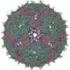
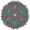
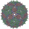
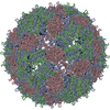
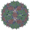
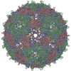
 Map data Map data | This is a surface-rendered view of a Bacteriophage MS2 virus-like particle composed of purified coat protein and genomic RNA | |||||||||
 Sample Sample |
| |||||||||
 Keywords Keywords |  Bacteriophage MS2 / virus assembly / quasi-equivalence / ssRNA virus Bacteriophage MS2 / virus assembly / quasi-equivalence / ssRNA virus | |||||||||
| Biological species |  Enterobacteria phage MS2 (virus) / Enterobacterio phage MS2 (virus) Enterobacteria phage MS2 (virus) / Enterobacterio phage MS2 (virus) | |||||||||
| Method | single particle reconstruction / cryo EM / Resolution: 32.0 Å | |||||||||
 Authors Authors | Rolfsson O / Toropova K / Ranson NA / Stockley PG | |||||||||
 Citation Citation | Journal: J Mol Biol / Year: 2010 Title: Mutually-induced conformational switching of RNA and coat protein underpins efficient assembly of a viral capsid. Authors: Óttar Rolfsson / Katerina Toropova / Neil A Ranson / Peter G Stockley /  Abstract: Single-stranded RNA viruses package their genomes into capsids enclosing fixed volumes. We assayed the ability of bacteriophage MS2 coat protein to package large, defined fragments of its genomic, ...Single-stranded RNA viruses package their genomes into capsids enclosing fixed volumes. We assayed the ability of bacteriophage MS2 coat protein to package large, defined fragments of its genomic, single-stranded RNA. We show that the efficiency of packaging into a T=3 capsid in vitro is inversely proportional to RNA length, implying that there is a free-energy barrier to be overcome during assembly. All the RNAs examined have greater solution persistence lengths than the internal diameter of the capsid into which they become packaged, suggesting that protein-mediated RNA compaction must occur during assembly. Binding ethidium bromide to one of these RNA fragments, which would be expected to reduce its flexibility, severely inhibited packaging, consistent with this idea. Cryo-EM structures of the capsids assembled in these experiments with the sub-genomic RNAs show a layer of RNA density beneath the coat protein shell but lack density for the inner RNA shell seen in the wild-type virion. The inner layer is restored when full-length virion RNA is used in the assembly reaction, implying that it becomes ordered only when the capsid is filled, presumably because of the effects of steric and/or electrostatic repulsions. The cryo-EM results explain the length dependence of packaging. In addition, they show that for the sub-genomic fragments the strongest ordered RNA density occurs below the coat protein dimers forming the icosahedral 5-fold axes of the capsid. There is little such density beneath the proteins at the 2-fold axes, consistent with our model in which coat protein dimers binding to RNA stem-loops located at sites throughout the genome leads to switching of their preferred conformations, thus regulating the placement of the quasi-conformers needed to build the T=3 capsid. The data are consistent with mutual chaperoning of both RNA and coat protein conformations, partially explaining the ability of such viruses to assemble so rapidly and accurately. | |||||||||
| History |
|
- Structure visualization
| Movie |
 Movie viewer Movie viewer |
|---|---|
| Structure viewer | EM map: SurfViewMolmilJmol/JSmol |
| Supplemental images |

- Downloads & links
-EMDB archive
| Map data | emd_1882.map.gz | 18.6 MB | EMDB map data format | |
|---|---|---|---|---|
| Header (meta data) | emd-1882-v30.xmlemd-1882.xml | 9.2 KB 9.2 KB | Display Display | EMDB header |
| Images | 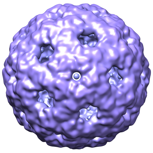 emd1882.jpg emd1882.jpg | 171.6 KB | ||
| Archive directory |  http://ftp.pdbj.org/pub/emdb/structures/EMD-1882ftp://ftp.pdbj.org/pub/emdb/structures/EMD-1882 http://ftp.pdbj.org/pub/emdb/structures/EMD-1882ftp://ftp.pdbj.org/pub/emdb/structures/EMD-1882 | HTTPS FTP |
-Related structure data
-Links
| EMDB pages | EMDB (EBI/PDBe) / EMDataResource |
|---|
-Map
| File | Download / File: emd_1882.map.gz / Format: CCP4 / Size: 29.8 MB / Type: IMAGE STORED AS FLOATING POINT NUMBER (4 BYTES) | ||||||||||||||||||||||||||||||||||||||||||||||||||||||||||||||||||||
|---|---|---|---|---|---|---|---|---|---|---|---|---|---|---|---|---|---|---|---|---|---|---|---|---|---|---|---|---|---|---|---|---|---|---|---|---|---|---|---|---|---|---|---|---|---|---|---|---|---|---|---|---|---|---|---|---|---|---|---|---|---|---|---|---|---|---|---|---|---|
| Annotation | This is a surface-rendered view of a Bacteriophage MS2 virus-like particle composed of purified coat protein and genomic RNA | ||||||||||||||||||||||||||||||||||||||||||||||||||||||||||||||||||||
| Voxel size | X=Y=Z: 1.87 Å | ||||||||||||||||||||||||||||||||||||||||||||||||||||||||||||||||||||
| Density |
| ||||||||||||||||||||||||||||||||||||||||||||||||||||||||||||||||||||
| Symmetry | Space group: 1 | ||||||||||||||||||||||||||||||||||||||||||||||||||||||||||||||||||||
| Details | EMDB XML:
CCP4 map header:
| ||||||||||||||||||||||||||||||||||||||||||||||||||||||||||||||||||||
-Supplemental data
- Sample components
-Entire : Bacteriophage MS2 virus-like particles assembled in vitro from pu...
| Entire | Name: Bacteriophage MS2 virus-like particles assembled in vitro from purified MS2 coat protein and genomic RNA |
|---|---|
| Components |
|
-Supramolecule #1000: Bacteriophage MS2 virus-like particles assembled in vitro from pu...
| Supramolecule | Name: Bacteriophage MS2 virus-like particles assembled in vitro from purified MS2 coat protein and genomic RNA type: sample / ID: 1000 / Oligomeric state: One capsid to one RNA / Number unique components: 2 |
|---|
-Supramolecule #1: Enterobacterio phage MS2
| Supramolecule | Name: Enterobacterio phage MS2 / type: virus / ID: 1 / Name.synonym: MS2 / NCBI-ID: 12022 / Sci species name: Enterobacterio phage MS2 / Virus type: VIRUS-LIKE PARTICLE / Virus isolate: STRAIN / Virus enveloped: No / Virus empty: No / Syn species name: MS2 |
|---|---|
| Host (natural) | Organism:  Escherichia coli (E. coli) / synonym: BACTERIA(EUBACTERIA) Escherichia coli (E. coli) / synonym: BACTERIA(EUBACTERIA) |
| Virus shell | Shell ID: 1 / Diameter: 280 Å / T number (triangulation number): 3 |
-Macromolecule #1: Single stranded RNA MS2 genome
| Macromolecule | Name: Single stranded RNA MS2 genome / type: rna / ID: 1 / Name.synonym: Single stranded RNA MS2 genome Details: Single stranded RNA MS2 genome was phenol-chloroform extracted from wild type MS2 phage Classification: OTHER / Structure: SINGLE STRANDED / Synthetic?: No |
|---|---|
| Source (natural) | Organism: Enterobacteria phage MS2 (virus) / synonym: MS2 |
-Experimental details
-Structure determination
| Method | cryo EM |
|---|---|
 Processing Processing | single particle reconstruction |
| Aggregation state | particle |
-Sample preparation
| Buffer | pH: 7.6 Details: 50mM Tris-acetate, 1mM magnesium acetate, 40mM ammonium acetate |
|---|---|
| Grid | Details: Quantifoil R2/1 200 mesh holly carbon grid |
| Vitrification | Cryogen name: ETHANE / Instrument: HOMEMADE PLUNGER Details: Vitrification instrument: Double-sided custom pneumatic blotter Method: 1.6s blot |
- Electron microscopy
| Microscope | FEI TECNAI F20 |
|---|---|
| Electron beam | Acceleration voltage: 200 kV / Electron source: FIELD EMISSION GUN |
| Electron optics | Calibrated magnification: 52750 / Illumination mode: FLOOD BEAM / Imaging mode: BRIGHT FIELDBright-field microscopy / Cs: 2.0 mm / Nominal magnification: 50000 |
| Sample stage | Specimen holder: Side entry / Specimen holder model: GATAN LIQUID NITROGEN |
| Image recording | Category: FILM / Film or detector model: KODAK SO-163 FILM / Digitization - Scanner: OTHER / Digitization - Sampling interval: 9.88 µm / Average electron dose: 18 e/Å2 / Bits/pixel: 16 |
| Experimental equipment |  Model: Tecnai F20 / Image courtesy: FEI Company |
-Image processing
| CTF correction | Details: Phase flip each particle |
|---|---|
| Final two d classification | Number classes: 19 |
| Final reconstruction | Applied symmetry - Point group: I (icosahedral) / Algorithm: OTHER / Resolution.type: BY AUTHOR / Resolution: 32.0 Å / Resolution method: FSC 0.5 CUT-OFF / Software - Name: Spider / Number images used: 80 |
