 Movie
Movie Controller

[English] 日本語
 Yorodumi
- EMDB-1867: Structure of the yeast eisosome core component Lsp1 filament boun... -
+ Open data

- Basic information
| Entry | Database: EMDB / ID: EMD-1867 | |||||||||
|---|---|---|---|---|---|---|---|---|---|---|
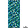
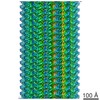
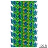
| Title | Structure of the yeast eisosome core component Lsp1 filament bound to a liposome membrane. | |||||||||
 Map data Map data | This is a map of Lsp1 filament bound to a liposome membrane with helical symmetry parameters turn -80.8 degrees, rise 4.9 A | |||||||||
 Sample Sample |
| |||||||||
| Biological species |  | |||||||||
| Method | helical reconstruction / cryo EM / Resolution: 31.0 Å | |||||||||
 Authors Authors | Karotki L / Huiskonen JT / Stefan CJ / Roth R / Surma MA / Aguilar PS / Krogan NJ / Emr SD / Heuser J / Gruenewald K / Walther TC | |||||||||
 Citation Citation | Journal: J Cell Biol / Year: 2011 Title: Eisosome proteins assemble into a membrane scaffold. Authors: Lena Karotki / Juha T Huiskonen / Christopher J Stefan / Natasza E Ziółkowska / Robyn Roth / Michal A Surma / Nevan J Krogan / Scott D Emr / John Heuser / Kay Grünewald / Tobias C Walther /  Abstract: Spatial organization of membranes into domains of distinct protein and lipid composition is a fundamental feature of biological systems. The plasma membrane is organized in such domains to ...Spatial organization of membranes into domains of distinct protein and lipid composition is a fundamental feature of biological systems. The plasma membrane is organized in such domains to efficiently orchestrate the many reactions occurring there simultaneously. Despite the almost universal presence of membrane domains, mechanisms of their formation are often unclear. Yeast cells feature prominent plasma membrane domain organization, which is at least partially mediated by eisosomes. Eisosomes are large protein complexes that are primarily composed of many subunits of two Bin-Amphiphysin-Rvs domain-containing proteins, Pil1 and Lsp1. In this paper, we show that these proteins self-assemble into higher-order structures and bind preferentially to phosphoinositide-containing membranes. Using a combination of electron microscopy approaches, we generate structural models of Pil1 and Lsp1 assemblies, which resemble eisosomes in cells. Our data suggest that the mechanism of membrane organization by eisosomes is mediated by self-assembly of its core components into a membrane-bound protein scaffold with lipid-binding specificity. | |||||||||
| History |
|
- Structure visualization
| Movie |
 Movie viewer Movie viewer |
|---|---|
| Structure viewer | EM map: SurfViewMolmilJmol/JSmol |
| Supplemental images |

- Downloads & links
-EMDB archive
| Map data | emd_1867.map.gz | 4.3 MB | EMDB map data format | |
|---|---|---|---|---|
| Header (meta data) | emd-1867-v30.xmlemd-1867.xml | 9.1 KB 9.1 KB | Display Display | EMDB header |
| Images | 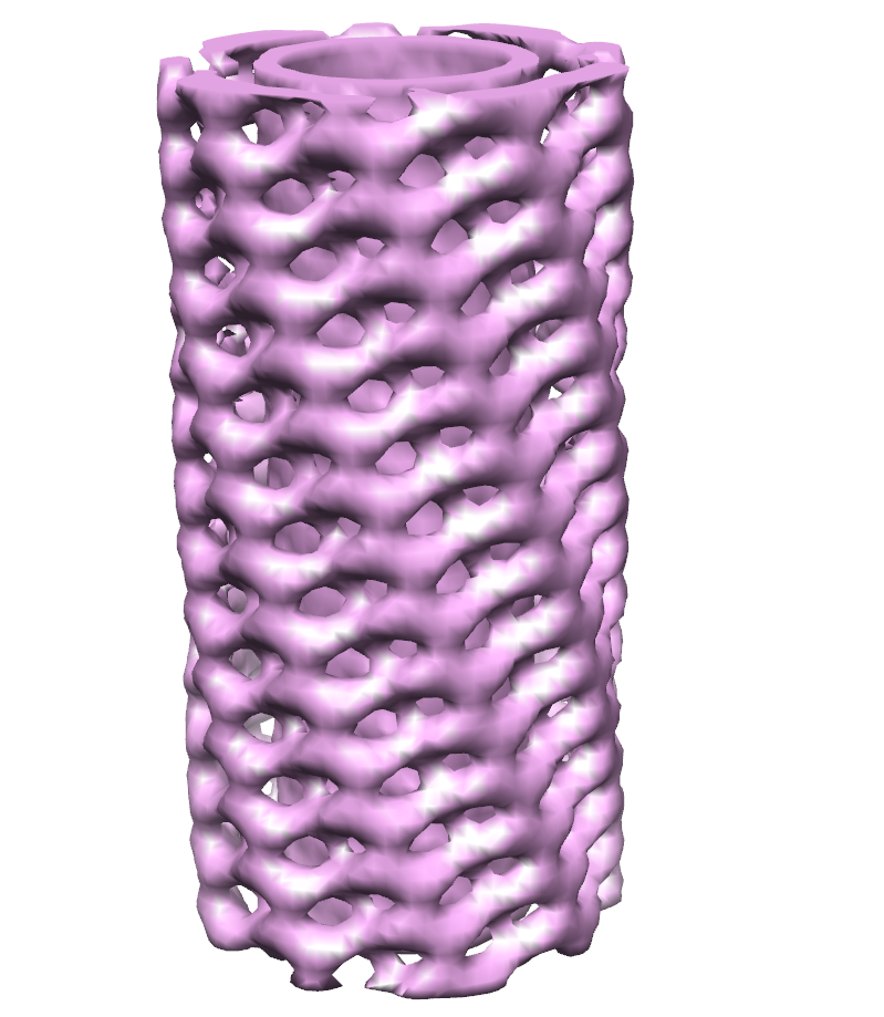 emd-1867.png emd-1867.png | 375.9 KB | ||
| Archive directory |  http://ftp.pdbj.org/pub/emdb/structures/EMD-1867ftp://ftp.pdbj.org/pub/emdb/structures/EMD-1867 http://ftp.pdbj.org/pub/emdb/structures/EMD-1867ftp://ftp.pdbj.org/pub/emdb/structures/EMD-1867 | HTTPS FTP |
-Validation report
| Summary document | emd_1867_validation.pdf.gz | 240.1 KB | Display | EMDB validaton report |
|---|---|---|---|---|
| Full document | emd_1867_full_validation.pdf.gz | 239.1 KB | Display | |
| Data in XML | emd_1867_validation.xml.gz | 5.8 KB | Display | |
| Arichive directory | https://ftp.pdbj.org/pub/emdb/validation_reports/EMD-1867ftp://ftp.pdbj.org/pub/emdb/validation_reports/EMD-1867 | HTTPS FTP |
-Related structure data
-Links
| EMDB pages | EMDB (EBI/PDBe) / EMDataResource |
|---|
-Map
| File | Download / File: emd_1867.map.gz / Format: CCP4 / Size: 12.6 MB / Type: IMAGE STORED AS FLOATING POINT NUMBER (4 BYTES) | ||||||||||||||||||||||||||||||||||||||||||||||||||||||||||||||||||||
|---|---|---|---|---|---|---|---|---|---|---|---|---|---|---|---|---|---|---|---|---|---|---|---|---|---|---|---|---|---|---|---|---|---|---|---|---|---|---|---|---|---|---|---|---|---|---|---|---|---|---|---|---|---|---|---|---|---|---|---|---|---|---|---|---|---|---|---|---|---|
| Annotation | This is a map of Lsp1 filament bound to a liposome membrane with helical symmetry parameters turn -80.8 degrees, rise 4.9 A | ||||||||||||||||||||||||||||||||||||||||||||||||||||||||||||||||||||
| Voxel size | X=Y=Z: 4.42 Å | ||||||||||||||||||||||||||||||||||||||||||||||||||||||||||||||||||||
| Density |
| ||||||||||||||||||||||||||||||||||||||||||||||||||||||||||||||||||||
| Symmetry | Space group: 1 | ||||||||||||||||||||||||||||||||||||||||||||||||||||||||||||||||||||
| Details | EMDB XML:
CCP4 map header:
| ||||||||||||||||||||||||||||||||||||||||||||||||||||||||||||||||||||
-Supplemental data
- Sample components
-Entire : Lsp1
| Entire | Name: Lsp1 |
|---|---|
| Components |
|
-Supramolecule #1000: Lsp1
| Supramolecule | Name: Lsp1 / type: sample / ID: 1000 / Oligomeric state: Helical multimer / Number unique components: 1 |
|---|
-Macromolecule #1: Yeast eisosome core component Lsp1 filament
| Macromolecule | Name: Yeast eisosome core component Lsp1 filament / type: protein_or_peptide / ID: 1 / Name.synonym: Lsp1 filament / Recombinant expression: Yes |
|---|---|
| Source (natural) | Organism: |
| Recombinant expression | Organism:  |
-Experimental details
-Structure determination
| Method | cryo EM |
|---|---|
 Processing Processing | helical reconstruction |
| Aggregation state | filament |
-Sample preparation
| Buffer | pH: 7.4 Details: 150mM KAcetate, 2mM MgAcetate, 20mM HEPES pH 7.4, 5% Glycerol |
|---|---|
| Grid | Details: Cflat CF-2/1-2C |
| Vitrification | Cryogen name: NITROGEN / Instrument: OTHER |
- Electron microscopy
| Microscope | FEI TECNAI F20 |
|---|---|
| Temperature | Average: 80 K |
| Details | Low dose imaging |
| Image recording | Category: CCD / Film or detector model: FEI EAGLE (4k x 4k) / Average electron dose: 20 e/Å2 |
| Electron beam | Acceleration voltage: 200 kV / Electron source:  FIELD EMISSION GUN FIELD EMISSION GUN |
| Electron optics | Calibrated magnification: 68180 / Illumination mode: FLOOD BEAM / Imaging mode: BRIGHT FIELD / Cs: 2.0 mm / Nominal magnification: 50000 |
| Sample stage | Specimen holder: Side entry / Specimen holder model: GATAN LIQUID NITROGEN |
| Experimental equipment |  Model: Tecnai F20 / Image courtesy: FEI Company |
-Image processing
| Final reconstruction | Applied symmetry - Helical parameters - Δz: 4.9 Å Applied symmetry - Helical parameters - Δ&Phi: 80.8 ° Applied symmetry - Helical parameters - Axial symmetry: C1 (asymmetric) Algorithm: OTHER / Resolution.type: BY AUTHOR / Resolution: 31.0 Å / Resolution method: FSC 0.5 CUT-OFF / Software - Name: IHRSR |
|---|---|
| CTF correction | Details: Phase flip for each particle |
