United Kingdom, France, United States, European Union, 10 items
Organization
Grant number
Country
Biotechnology and Biological Sciences Research Council (BBSRC)
EP/V051474/1
United Kingdom
Medical Research Council (MRC, United Kingdom)
MC UP 1201/31
United Kingdom
Human Frontier Science Program (HFSP)
RGY0074/2021
France
Engineering and Physical Sciences Research Council
EP/V026623/1
United Kingdom
The Vallee Foundation Inc.
United States
European Molecular Biology Organization (EMBO)
European Union
Leverhulme Trust
United Kingdom
The Lister Institute of Preventive Medicine
United Kingdom
Wellcome Trust
104633/Z/14/Z
United Kingdom
Royal Society
NIF/R1/192285
United Kingdom
Citation
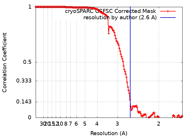
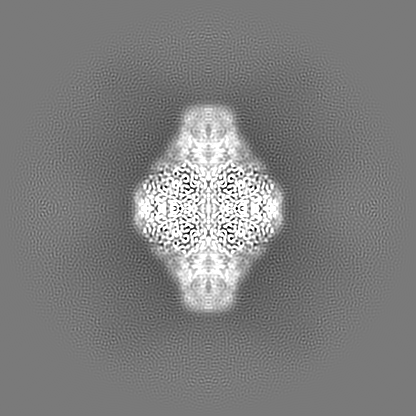
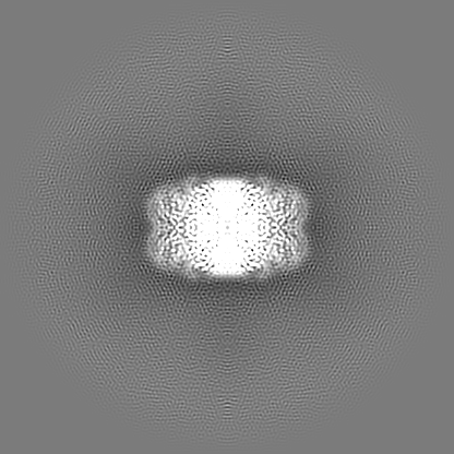
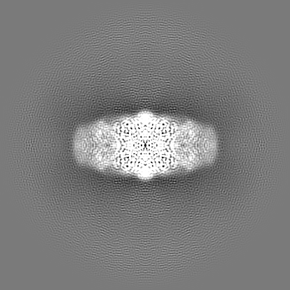
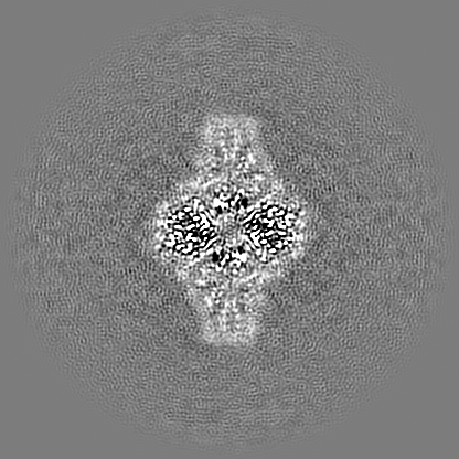
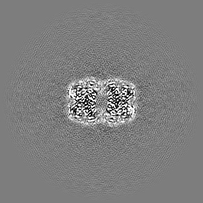
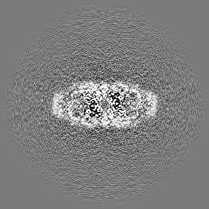
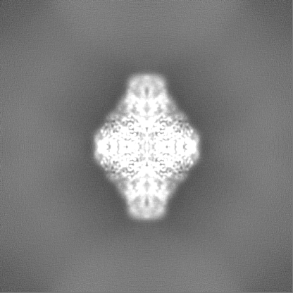
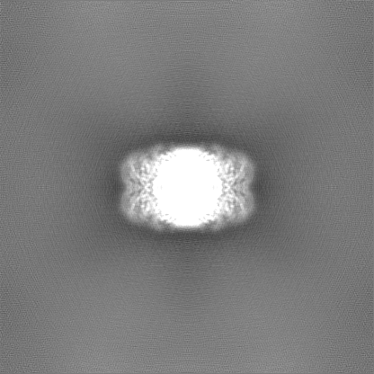
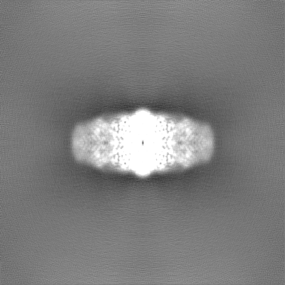
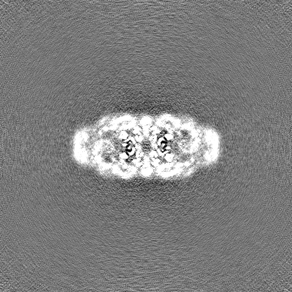
Journal: Sci Adv / Year: 2024 Title: Cryo-EM of soft-landed β-galactosidase: Gas-phase and native structures are remarkably similar. Authors: Tim K Esser / Jan Böhning / Alpcan Önür / Dinesh K Chinthapalli / Lukas Eriksson / Marko Grabarics / Paul Fremdling / Albert Konijnenberg / Alexander Makarov / Aurelien Botman / Christine ...Authors: Tim K Esser / Jan Böhning / Alpcan Önür / Dinesh K Chinthapalli / Lukas Eriksson / Marko Grabarics / Paul Fremdling / Albert Konijnenberg / Alexander Makarov / Aurelien Botman / Christine Peter / Justin L P Benesch / Carol V Robinson / Joseph Gault / Lindsay Baker / Tanmay A M Bharat / Stephan Rauschenbach / Abstract: Native mass spectrometry (MS) has become widely accepted in structural biology, providing information on stoichiometry, interactions, homogeneity, and shape of protein complexes. Yet, the fundamental ...Native mass spectrometry (MS) has become widely accepted in structural biology, providing information on stoichiometry, interactions, homogeneity, and shape of protein complexes. Yet, the fundamental assumption that proteins inside the mass spectrometer retain a structure faithful to native proteins in solution remains a matter of intense debate. Here, we reveal the gas-phase structure of β-galactosidase using single-particle cryo-electron microscopy (cryo-EM) down to 2.6-Å resolution, enabled by soft landing of mass-selected protein complexes onto cold transmission electron microscopy (TEM) grids followed by in situ ice coating. We find that large parts of the secondary and tertiary structure are retained from the solution. Dehydration-driven subunit reorientation leads to consistent compaction in the gas phase. By providing a direct link between high-resolution imaging and the capability to handle and select protein complexes that behave problematically in conventional sample preparation, the approach has the potential to expand the scope of both native mass spectrometry and cryo-EM.
In the structure databanks used in Yorodumi, some data are registered as the other names, "COVID-19 virus" and "2019-nCoV". Here are the details of the virus and the list of structure data.
Jan 31, 2019. EMDB accession codes are about to change! (news from PDBe EMDB page)
EMDB accession codes are about to change! (news from PDBe EMDB page)
The allocation of 4 digits for EMDB accession codes will soon come to an end. Whilst these codes will remain in use, new EMDB accession codes will include an additional digit and will expand incrementally as the available range of codes is exhausted. The current 4-digit format prefixed with “EMD-” (i.e. EMD-XXXX) will advance to a 5-digit format (i.e. EMD-XXXXX), and so on. It is currently estimated that the 4-digit codes will be depleted around Spring 2019, at which point the 5-digit format will come into force.
The EM Navigator/Yorodumi systems omit the EMD- prefix.
Related info.:Q: What is EMD? / ID/Accession-code notation in Yorodumi/EM Navigator
Yorodumi is a browser for structure data from EMDB, PDB, SASBDB, etc.
This page is also the successor to EM Navigator detail page, and also detail information page/front-end page for Omokage search.
The word "yorodu" (or yorozu) is an old Japanese word meaning "ten thousand". "mi" (miru) is to see.
Related info.:EMDB / PDB / SASBDB / Comparison of 3 databanks / Yorodumi Search / Aug 31, 2016. New EM Navigator & Yorodumi / Yorodumi Papers / Jmol/JSmol / Function and homology information / Changes in new EM Navigator and Yorodumi
 Movie
Movie Controller


 Open data
 Basic information
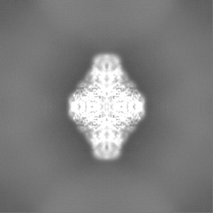
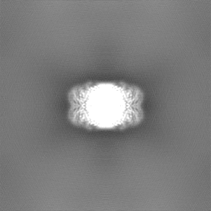
 Map data
 Sample
 Keywords
 Lactase /
  Function and homology information
 Authors
 United Kingdom,
  France,
  United States, European Union, 10 items
  Citation

 Structure visualization
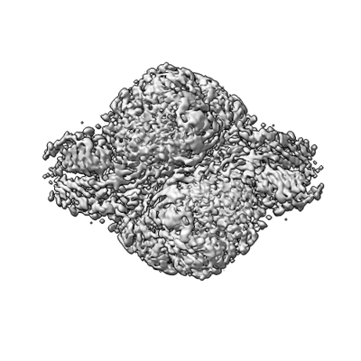
 Downloads & links
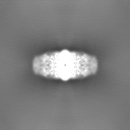
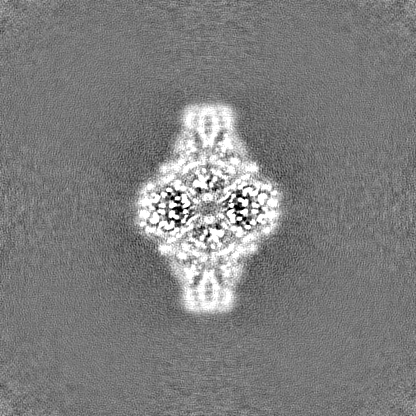
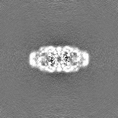
 emd_18244.png
 http://ftp.pdbj.org/pub/emdb/structures/EMD-18244
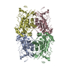


 Z
Z Y
Y X
X


 Sample components
 Processing
 Electron microscopy