 Movie
Movie Controller

[English] 日本語
 Yorodumi
- EMDB-1774: Understanding Ribosome Assembly: The Structure of in vivo Assembl... -
+ Open data

- Basic information
| Entry | Database: EMDB / ID: EMD-1774 | |||||||||
|---|---|---|---|---|---|---|---|---|---|---|
| Title | Understanding Ribosome Assembly: The Structure of in vivo Assembled Immature 30S Subunits Revealed by Cryo-Electron Microscopy | |||||||||
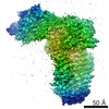
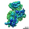
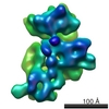
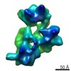
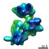
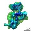
 Map data Map data | Surface rendering of the immature 30S ribosomal subunit from YjeQ-depleted E.coli cells. | |||||||||
 Sample Sample |
| |||||||||
 Keywords Keywords | 3D Structure of Immature 30S Subunit | |||||||||
| Biological species |  | |||||||||
| Method | single particle reconstruction / cryo EM / Resolution: 11.6 Å | |||||||||
 Authors Authors | Jomaa A / Stewart G / Benito JM / Zielke R / Campbell T / Maddock J / Brown E / Ortega J | |||||||||
 Citation Citation | Journal: RNA / Year: 2011 Title: Understanding ribosome assembly: the structure of in vivo assembled immature 30S subunits revealed by cryo-electron microscopy. Authors: Ahmad Jomaa / Geordie Stewart / Jaime Martín-Benito / Ryszard Zielke / Tracey L Campbell / Janine R Maddock / Eric D Brown / Joaquin Ortega /  Abstract: Four decades after early in vitro assembly studies demonstrated that ribosome assembly is a controlled process, our understanding of ribosome assembly is still incomplete. Just as structure ...Four decades after early in vitro assembly studies demonstrated that ribosome assembly is a controlled process, our understanding of ribosome assembly is still incomplete. Just as structure determination has been so important to understanding ribosome function, so too will it be critical to sorting out the assembly process. Here, we used a viable deletion in the yjeQ gene, a recognized ribosome assembly factor, to isolate and structurally characterize immature 30S subunits assembled in vivo. These small ribosome subunits contained unprocessed 17S rRNA and lacked some late ribosomal proteins. Cryo-electron microscopy reconstructions revealed that the presence of precursor sequences in the rRNA induces a severe distortion in the 3' minor domain of the subunit involved in the decoding of mRNA and interaction with the large ribosome subunit. These findings suggest that rRNA processing events induce key local conformational changes directing the structure toward the mature assembly. We concluded that rRNA processing, folding, and the entry of tertiary r-proteins are interdependent events in the late stages of 30S subunit assembly. In addition, we demonstrate how studies of emerging assembly factors in ribosome biogenesis can help to elucidate the path of subunit assembly in vivo. | |||||||||
| History |
|
- Structure visualization
| Movie |
 Movie viewer Movie viewer |
|---|---|
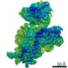
| Structure viewer | EM map: SurfViewMolmilJmol/JSmol |
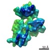
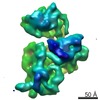
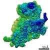
| Supplemental images |

- Downloads & links
-EMDB archive
| Map data | emd_1774.map.gz | 7.4 MB | EMDB map data format | |
|---|---|---|---|---|
| Header (meta data) | emd-1774-v30.xmlemd-1774.xml | 10.2 KB 10.2 KB | Display Display | EMDB header |
| Images | 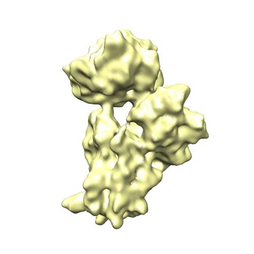 EMD-1774.jpg EMD-1774.jpg | 38.1 KB | ||
| Archive directory |  http://ftp.pdbj.org/pub/emdb/structures/EMD-1774ftp://ftp.pdbj.org/pub/emdb/structures/EMD-1774 http://ftp.pdbj.org/pub/emdb/structures/EMD-1774ftp://ftp.pdbj.org/pub/emdb/structures/EMD-1774 | HTTPS FTP |
-Validation report
| Summary document | emd_1774_validation.pdf.gz | 233.1 KB | Display | EMDB validaton report |
|---|---|---|---|---|
| Full document | emd_1774_full_validation.pdf.gz | 232.2 KB | Display | |
| Data in XML | emd_1774_validation.xml.gz | 5.5 KB | Display | |
| Arichive directory | https://ftp.pdbj.org/pub/emdb/validation_reports/EMD-1774ftp://ftp.pdbj.org/pub/emdb/validation_reports/EMD-1774 | HTTPS FTP |
-Related structure data

-Links
| EMDB pages | EMDB (EBI/PDBe) / EMDataResource |
|---|---|
| Related items in Molecule of the Month |

-Map
| File | Download / File: emd_1774.map.gz / Format: CCP4 / Size: 7.8 MB / Type: IMAGE STORED AS FLOATING POINT NUMBER (4 BYTES) | ||||||||||||||||||||||||||||||||||||||||||||||||||||||||||||||||||||
|---|---|---|---|---|---|---|---|---|---|---|---|---|---|---|---|---|---|---|---|---|---|---|---|---|---|---|---|---|---|---|---|---|---|---|---|---|---|---|---|---|---|---|---|---|---|---|---|---|---|---|---|---|---|---|---|---|---|---|---|---|---|---|---|---|---|---|---|---|---|
| Annotation | Surface rendering of the immature 30S ribosomal subunit from YjeQ-depleted E.coli cells. | ||||||||||||||||||||||||||||||||||||||||||||||||||||||||||||||||||||
| Voxel size | X=Y=Z: 2.54 Å | ||||||||||||||||||||||||||||||||||||||||||||||||||||||||||||||||||||
| Density |
| ||||||||||||||||||||||||||||||||||||||||||||||||||||||||||||||||||||
| Symmetry | Space group: 1 | ||||||||||||||||||||||||||||||||||||||||||||||||||||||||||||||||||||
| Details | EMDB XML:
CCP4 map header:
| ||||||||||||||||||||||||||||||||||||||||||||||||||||||||||||||||||||
-Supplemental data
- Sample components
-Entire : Reconstruction of the immature 30S ribosomal subunit purified fro...
| Entire | Name: Reconstruction of the immature 30S ribosomal subunit purified from yjeq-deficient E.coli strains. |
|---|---|
| Components |
|
-Supramolecule #1000: Reconstruction of the immature 30S ribosomal subunit purified fro...
| Supramolecule | Name: Reconstruction of the immature 30S ribosomal subunit purified from yjeq-deficient E.coli strains. type: sample / ID: 1000 Details: The sample was thawed from -80 degrees Celsius and diluted to a concentration of 58 nM. Sample was then applied on holey carbon grids with an additional thin layer of carbon (5-10 nm) and ...Details: The sample was thawed from -80 degrees Celsius and diluted to a concentration of 58 nM. Sample was then applied on holey carbon grids with an additional thin layer of carbon (5-10 nm) and plunged in liquid ethane at liquid nitrogen temperature. Number unique components: 1 |
|---|---|
| Molecular weight | Theoretical: 800 KDa |
-Supramolecule #1: Small Ribosomal Subunit
| Supramolecule | Name: Small Ribosomal Subunit / type: complex / ID: 1 / Name.synonym: 30S / Recombinant expression: No / Ribosome-details: ribosome-prokaryote: SSU 30S |
|---|---|
| Source (natural) | Organism: |
| Molecular weight | Theoretical: 850 KDa |
-Experimental details
-Structure determination
| Method | cryo EM |
|---|---|
 Processing Processing | single particle reconstruction |
| Aggregation state | particle |
-Sample preparation
| Concentration | 4.7 mg/mL |
|---|---|
| Buffer | pH: 7.5 Details: 10 mM Tris-HCl, 10 mM magnesium acetate, 60 mM NH4Cl, 3 mM 2-mercaptoethanol |
| Grid | Details: 400 mesh Copper Grid |
| Vitrification | Cryogen name: ETHANE / Chamber humidity: 100 % / Chamber temperature: 93 K / Instrument: OTHER / Details: Vitrification instrument: FEI vitrobot / Method: Blot for 7 seconds twice before plunging |
- Electron microscopy
| Microscope | JEOL 2010F |
|---|---|
| Temperature | Min: 93 K / Max: 93 K / Average: 93 K |
| Date | Oct 16, 2008 |
| Image recording | Category: FILM / Film or detector model: KODAK SO-163 FILM / Digitization - Scanner: NIKON SUPER COOLSCAN 9000 / Digitization - Sampling interval: 12.7 µm / Number real images: 72 / Average electron dose: 10 e/Å2 / Bits/pixel: 16 |
| Electron beam | Acceleration voltage: 200 kV / Electron source:  FIELD EMISSION GUN FIELD EMISSION GUN |
| Electron optics | Calibrated magnification: 50000 / Illumination mode: FLOOD BEAM / Imaging mode: BRIGHT FIELD / Cs: 1.0 mm / Nominal defocus max: 3.9 µm / Nominal defocus min: 0.65 µm / Nominal magnification: 50000 |
| Sample stage | Specimen holder: Side entry Liquid Nitrogen-Cooled Cryo Specimen holder.This holder operates in the temperature range from -175 C to ambient. Specimen holder model: GATAN LIQUID NITROGEN |
-Image processing
| Details | Particles were picked using BOXER software implemented in EMAN. |
|---|---|
| CTF correction | Details: Each Micrograph |
| Final reconstruction | Applied symmetry - Point group: C1 (asymmetric) / Algorithm: OTHER / Resolution.type: BY AUTHOR / Resolution: 11.6 Å / Resolution method: FSC 0.5 CUT-OFF / Software - Name: Xmipp / Number images used: 59054 |
-Atomic model buiding 1
| Initial model | PDB ID:  2z4k |
|---|---|
| Software | Name: Situs |
| Details | Protocol: Rigid Body |
| Refinement | Space: REAL / Protocol: RIGID BODY FIT |
