 Movie
Movie Controller

[English] 日本語
 Yorodumi
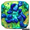
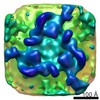
- EMDB-1704: Sub-tomogram average of Tula Hantavirus membrane glycoprotein lattice -
+ Open data

- Basic information
| Entry | Database: EMDB / ID: EMD-1704 | |||||||||
|---|---|---|---|---|---|---|---|---|---|---|
| Title | Sub-tomogram average of Tula Hantavirus membrane glycoprotein lattice | |||||||||
 Map data Map data | Sub-tomogram average of Tula Hantavirus membrane glycoprotein lattice | |||||||||
 Sample Sample |
| |||||||||
 Keywords Keywords | Tula / hantavirus / bunyavirus / bunyaviridae / glycoprotein N / glycoprotein G / membrane | |||||||||
| Biological species |  Hantavirus Hantavirus | |||||||||
| Method | subtomogram averaging / cryo EM / Resolution: 36.0 Å | |||||||||
 Authors Authors | Huiskonen JT / Hepojoki J / Laurinmaki P / Vaheri A / Lankinen H / Butcher SJ / Grunewald K | |||||||||
 Citation Citation | Journal: J Virol / Year: 2010 Title: Electron cryotomography of Tula hantavirus suggests a unique assembly paradigm for enveloped viruses. Authors: Juha T Huiskonen / Jussi Hepojoki / Pasi Laurinmäki / Antti Vaheri / Hilkka Lankinen / Sarah J Butcher / Kay Grünewald /  Abstract: Hantaviruses (family Bunyaviridae) are rodent-borne emerging viruses that cause a serious, worldwide threat to human health. Hantavirus diseases include hemorrhagic fever with renal syndrome and ...Hantaviruses (family Bunyaviridae) are rodent-borne emerging viruses that cause a serious, worldwide threat to human health. Hantavirus diseases include hemorrhagic fever with renal syndrome and hantavirus cardiopulmonary syndrome. Virions are enveloped and contain a tripartite single-stranded negative-sense RNA genome. Two types of glycoproteins, G(N) and G(C), are embedded in the viral membrane and form protrusions, or "spikes." The membrane encloses a ribonucleoprotein core, which consists of the RNA segments, the nucleocapsid protein, and the RNA-dependent RNA polymerase. Detailed information on hantavirus virion structure and glycoprotein spike composition is scarce. Here, we have studied the structures of Tula hantavirus virions using electron cryomicroscopy and tomography. Three-dimensional density maps show how the hantavirus surface glycoproteins, membrane, and ribonucleoprotein are organized. The structure of the G(N)-G(C) spike complex was solved to 3.6-nm resolution by averaging tomographic subvolumes. Each spike complex is a square-shaped assembly with 4-fold symmetry. Spike complexes formed ordered patches on the viral membrane by means of specific lateral interactions. These interactions may be sufficient for creating membrane curvature during virus budding. In conclusion, the structure and assembly principles of Tula hantavirus exemplify a unique assembly paradigm for enveloped viruses. | |||||||||
| History |
|
- Structure visualization
| Movie |
 Movie viewer Movie viewer |
|---|---|
| Structure viewer | EM map: SurfViewMolmilJmol/JSmol |
| Supplemental images |

- Downloads & links
-EMDB archive
| Map data | emd_1704.map.gz | 2.4 MB | EMDB map data format | |
|---|---|---|---|---|
| Header (meta data) | emd-1704-v30.xmlemd-1704.xml | 9.7 KB 9.7 KB | Display Display | EMDB header |
| Images | 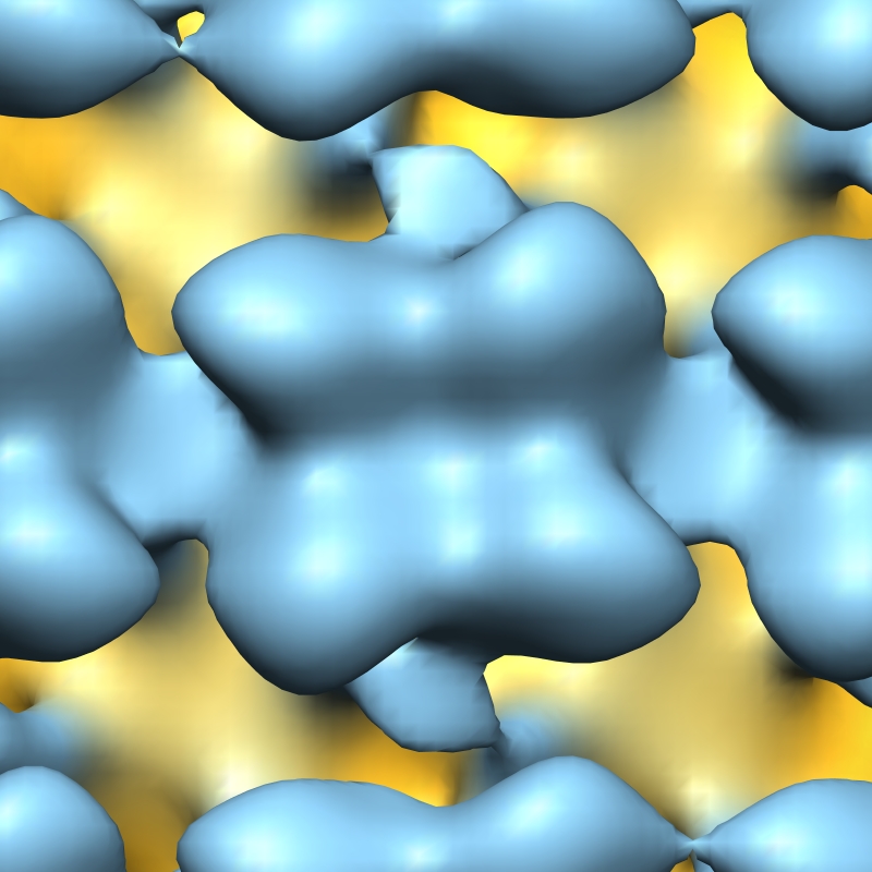 1704.jpg 1704.jpg | 241.2 KB | ||
| Archive directory |  http://ftp.pdbj.org/pub/emdb/structures/EMD-1704ftp://ftp.pdbj.org/pub/emdb/structures/EMD-1704 http://ftp.pdbj.org/pub/emdb/structures/EMD-1704ftp://ftp.pdbj.org/pub/emdb/structures/EMD-1704 | HTTPS FTP |
-Validation report
| Summary document | emd_1704_validation.pdf.gz | 224.5 KB | Display | EMDB validaton report |
|---|---|---|---|---|
| Full document | emd_1704_full_validation.pdf.gz | 223.7 KB | Display | |
| Data in XML | emd_1704_validation.xml.gz | 5.3 KB | Display | |
| Arichive directory | https://ftp.pdbj.org/pub/emdb/validation_reports/EMD-1704ftp://ftp.pdbj.org/pub/emdb/validation_reports/EMD-1704 | HTTPS FTP |
-Related structure data
| Similar structure data |
|---|

-Links
| EMDB pages | EMDB (EBI/PDBe) / EMDataResource |
|---|
-Map
| File | Download / File: emd_1704.map.gz / Format: CCP4 / Size: 2.7 MB / Type: IMAGE STORED AS FLOATING POINT NUMBER (4 BYTES) | ||||||||||||||||||||||||||||||||||||||||||||||||||||||||||||||||||||
|---|---|---|---|---|---|---|---|---|---|---|---|---|---|---|---|---|---|---|---|---|---|---|---|---|---|---|---|---|---|---|---|---|---|---|---|---|---|---|---|---|---|---|---|---|---|---|---|---|---|---|---|---|---|---|---|---|---|---|---|---|---|---|---|---|---|---|---|---|---|
| Annotation | Sub-tomogram average of Tula Hantavirus membrane glycoprotein lattice | ||||||||||||||||||||||||||||||||||||||||||||||||||||||||||||||||||||
| Voxel size | X=Y=Z: 4.46 Å | ||||||||||||||||||||||||||||||||||||||||||||||||||||||||||||||||||||
| Density |
| ||||||||||||||||||||||||||||||||||||||||||||||||||||||||||||||||||||
| Symmetry | Space group: 1 | ||||||||||||||||||||||||||||||||||||||||||||||||||||||||||||||||||||
| Details | EMDB XML:
CCP4 map header:
| ||||||||||||||||||||||||||||||||||||||||||||||||||||||||||||||||||||
-Supplemental data
- Sample components
-Entire : Tula hantavirus
| Entire | Name: Tula hantavirus |
|---|---|
| Components |
|
-Supramolecule #1000: Tula hantavirus
| Supramolecule | Name: Tula hantavirus / type: sample / ID: 1000 / Number unique components: 1 |
|---|
-Supramolecule #1: Hantavirus
| Supramolecule | Name: Hantavirus / type: virus / ID: 1 / Name.synonym: Tula hantavirus / NCBI-ID: 11598 / Sci species name: Hantavirus / Database: NCBI / Virus type: VIRION / Virus isolate: SPECIES / Virus enveloped: Yes / Virus empty: No / Syn species name: Tula hantavirus |
|---|---|
| Host (natural) | Organism:  Homo sapiens (human) / synonym: VERTEBRATES Homo sapiens (human) / synonym: VERTEBRATES |
| Virus shell | Shell ID: 1 / Name: Glycoprotein shell |
-Experimental details
-Structure determination
| Method | cryo EM |
|---|---|
 Processing Processing | subtomogram averaging |
-Sample preparation
| Buffer | pH: 7.4 / Details: 10 mM HEPES, 135 mM NaCl |
|---|---|
| Grid | Details: Cflat R2/2 |
| Vitrification | Cryogen name: ETHANE / Chamber temperature: 77 K / Instrument: HOMEMADE PLUNGER / Details: Vitrification instrument: MPI custom plunger / Method: Blot 4 seconds before plunging |
- Electron microscopy
| Microscope | FEI POLARA 300 |
|---|---|
| Temperature | Average: 77 K |
| Specialist optics | Energy filter - Name: GIF 2002 / Energy filter - Lower energy threshold: 0.0 eV / Energy filter - Upper energy threshold: 10.0 eV |
| Image recording | Category: CCD / Film or detector model: GENERIC CCD / Digitization - Sampling interval: 30 µm / Average electron dose: 100 e/Å2 / Bits/pixel: 8 |
| Electron beam | Acceleration voltage: 300 kV / Electron source:  FIELD EMISSION GUN FIELD EMISSION GUN |
| Electron optics | Calibrated magnification: 67300 / Illumination mode: FLOOD BEAM / Imaging mode: BRIGHT FIELD / Cs: 2.0 mm / Nominal defocus max: 6.0 µm / Nominal defocus min: 5.0 µm / Nominal magnification: 50000 |
| Sample stage | Specimen holder: Polara / Specimen holder model: OTHER / Tilt series - Axis1 - Min angle: -60 ° / Tilt series - Axis1 - Max angle: 60 ° |
| Experimental equipment |  Model: Tecnai Polara / Image courtesy: FEI Company |
-Image processing
| Details | Average number of tilts used in the 3D reconstructions: 61. Average tomographic tilt angle increment: 2. |
|---|---|
| Final reconstruction | Algorithm: OTHER / Resolution.type: BY AUTHOR / Resolution: 36.0 Å / Resolution method: FSC 0.5 CUT-OFF / Software - Name: Bsoft, jsubtomo Details: Sub-tomogram average calculated using 2,353 aligned spikes |
