 Movie
Movie Controller

[English] 日本語
 Yorodumi
- EMDB-1583: 3D structure of the C3bB complex provides insights into the activ... -
+ Open data

- Basic information
| Entry | Database: EMDB / ID: EMD-1583 | |||||||||
|---|---|---|---|---|---|---|---|---|---|---|
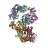
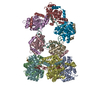
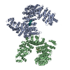
| Title | 3D structure of the C3bB complex provides insights into the activation and regulation of the complement alternative pathway convertase | |||||||||
 Map data Map data | Structure of the C3bB pro-convertase. | |||||||||
 Sample Sample |
| |||||||||
 Keywords Keywords | C3bB / complement alternative pathway | |||||||||
| Biological species |   Homo sapiens (human) Homo sapiens (human) | |||||||||
| Method | single particle reconstruction / negative staining / Resolution: 28.0 Å | |||||||||
 Authors Authors | Torreira E / Tortajada A / Montes T / Rodriguez de Cordoba S / Llorca O | |||||||||
 Citation Citation | Journal: Proc Natl Acad Sci U S A / Year: 2009 Title: 3D structure of the C3bB complex provides insights into the activation and regulation of the complement alternative pathway convertase. Authors: Eva Torreira / Agustín Tortajada / Tamara Montes / Santiago Rodríguez de Córdoba / Oscar Llorca /  Abstract: Generation of the alternative pathway C3-convertase, the central amplification enzyme of the complement cascade, initiates by the binding of factor B (fB) to C3b to form the proconvertase, C3bB. C3bB ...Generation of the alternative pathway C3-convertase, the central amplification enzyme of the complement cascade, initiates by the binding of factor B (fB) to C3b to form the proconvertase, C3bB. C3bB is subsequently cleaved by factor D (fD) at a single site in fB, producing Ba and Bb fragments. Ba dissociates from the complex, while Bb remains bound to C3b, forming the active alternative pathway convertase, C3bBb. Using single-particle electron microscopy we have determined the 3-dimensional structures of the C3bB and the C3bBb complexes at approximately 27A resolution. The C3bB structure shows that fB undergoes a dramatic conformational change upon binding to C3b. However, the C3b-bound fB structure was easily interpreted after independently fitting the atomic structures of the isolated Bb and Ba fragments. Interestingly, the divalent cation-binding site in the von Willebrand type A domain in Bb faces the C345C domain of C3b, whereas the serine-protease domain of Bb points outwards. The structure also shows that the Ba fragment interacts with C3b separately from Bb at the level of the alpha'NT and CUB domains. Within this conformation, the long and flexible linker between Bb and Ba is likely exposed and accessible for cleavage by fD to form the active convertase, C3bBb. The architecture of the C3bB and C3bBb complexes reveals that C3b could promote cleavage and activation of fB by actively displacing the Ba domain from the von Willebrand type A domain in free fB. These structures provide a structural basis to understand fundamental aspects of the activation and regulation of the alternative pathway C3-convertase. | |||||||||
| History |
|
- Structure visualization
| Movie |
 Movie viewer Movie viewer |
|---|---|
| Structure viewer | EM map: SurfViewMolmilJmol/JSmol |
| Supplemental images |

- Downloads & links
-EMDB archive
| Map data | emd_1583.map.gz | 139.6 KB | EMDB map data format | |
|---|---|---|---|---|
| Header (meta data) | emd-1583-v30.xmlemd-1583.xml | 8.1 KB 8.1 KB | Display Display | EMDB header |
| Images |  1583.gif 1583.gif | 55.4 KB | ||
| Archive directory |  http://ftp.pdbj.org/pub/emdb/structures/EMD-1583ftp://ftp.pdbj.org/pub/emdb/structures/EMD-1583 http://ftp.pdbj.org/pub/emdb/structures/EMD-1583ftp://ftp.pdbj.org/pub/emdb/structures/EMD-1583 | HTTPS FTP |
-Related structure data
| Similar structure data |
|---|


-Links
| EMDB pages | EMDB (EBI/PDBe) / EMDataResource |
|---|
-Map
| File | Download / File: emd_1583.map.gz / Format: CCP4 / Size: 1.4 MB / Type: IMAGE STORED AS FLOATING POINT NUMBER (4 BYTES) | ||||||||||||||||||||||||||||||||||||||||||||||||||||||||||||||||||||
|---|---|---|---|---|---|---|---|---|---|---|---|---|---|---|---|---|---|---|---|---|---|---|---|---|---|---|---|---|---|---|---|---|---|---|---|---|---|---|---|---|---|---|---|---|---|---|---|---|---|---|---|---|---|---|---|---|---|---|---|---|---|---|---|---|---|---|---|---|---|
| Annotation | Structure of the C3bB pro-convertase. | ||||||||||||||||||||||||||||||||||||||||||||||||||||||||||||||||||||
| Voxel size | X=Y=Z: 4.2 Å | ||||||||||||||||||||||||||||||||||||||||||||||||||||||||||||||||||||
| Density |
| ||||||||||||||||||||||||||||||||||||||||||||||||||||||||||||||||||||
| Symmetry | Space group: 1 | ||||||||||||||||||||||||||||||||||||||||||||||||||||||||||||||||||||
| Details | EMDB XML:
CCP4 map header:
| ||||||||||||||||||||||||||||||||||||||||||||||||||||||||||||||||||||
-Supplemental data
- Sample components
-Entire : C3bB
| Entire | Name: C3bB |
|---|---|
| Components |
|
-Supramolecule #1000: C3bB
| Supramolecule | Name: C3bB / type: sample / ID: 1000 Oligomeric state: One monomer of C3b binds to one monomer of Fb Number unique components: 2 |
|---|---|
| Molecular weight | Experimental: 270 KDa / Theoretical: 270 KDa |
-Macromolecule #1: C3bB
| Macromolecule | Name: C3bB / type: protein_or_peptide / ID: 1 / Name.synonym: complement pro-convertase / Number of copies: 1 / Oligomeric state: dimer / Recombinant expression: No |
|---|---|
| Source (natural) | Organism: Homo sapiens (human) / synonym: Human |
-Experimental details
-Structure determination
| Method | negative staining |
|---|---|
 Processing Processing | single particle reconstruction |
| Aggregation state | particle |
-Sample preparation
| Buffer | Details: 25mM TrisHCl pH8.0, 50mM NaCl, 5 mM NiCl2 |
|---|---|
| Staining | Type: NEGATIVE / Details: 1% uranyl formate |
| Grid | Details: 400 mesh Copper/Palladium grid |
| Vitrification | Cryogen name: NONE / Instrument: OTHER |
- Electron microscopy
| Microscope | JEOL 1230 |
|---|---|
| Electron beam | Acceleration voltage: 100 kV / Electron source: TUNGSTEN HAIRPIN |
| Electron optics | Illumination mode: FLOOD BEAM / Imaging mode: BRIGHT FIELDBright-field microscopy / Cs: 2.9 mm / Nominal magnification: 50000 |
| Sample stage | Specimen holder: Eucentric / Specimen holder model: OTHER / Tilt angle max: 35 |
| Image recording | Category: FILM / Film or detector model: KODAK SO-163 FILM / Digitization - Scanner: OTHER / Digitization - Sampling interval: 10.5 µm Details: images scanned with a MINOLTA Dimage Scan Multi Pro scanner at 2400 dpi and averaged to a final 4.2 angstroms pixel at the specimen Bits/pixel: 16 |
| Tilt angle min | 0 |
-Image processing
| Final reconstruction | Applied symmetry - Point group: C1 (asymmetric) / Resolution.type: BY AUTHOR / Resolution: 28.0 Å / Resolution method: FSC 0.5 CUT-OFF / Number images used: 6000 |
|---|
-Atomic model buiding 1
| Initial model | PDB ID: |
|---|---|
| Refinement | Space: RECIPROCAL |

