 Movie
Movie Controller

[English] 日本語
 Yorodumi
- EMDB-1575: C-terminal domain of adenovirus serotype 5 protein IX assemble in... -
+ Open data

- Basic information
| Entry | Database: EMDB / ID: EMD-1575 | |||||||||
|---|---|---|---|---|---|---|---|---|---|---|
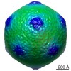
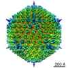
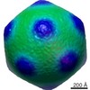
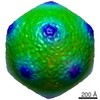
| Title | C-terminal domain of adenovirus serotype 5 protein IX assemble into an anti-parallel structure | |||||||||
 Map data Map data | Adenovirus sy12 with anti-sy12 fab | |||||||||
 Sample Sample |
| |||||||||
 Keywords Keywords |  adenovirus / proteinIX / Fab adenovirus / proteinIX / Fab | |||||||||
| Biological species |   Human adenovirus 5 Human adenovirus 5 | |||||||||
| Method | single particle reconstruction / cryo EM / negative staining / Resolution: 22.0 Å | |||||||||
 Authors Authors | Fabry CMS / Rosa-Calatrava M / Moriscot C / Ruigrok RWH / Boulanger P / Schoehn G | |||||||||
 Citation Citation | Journal: J Virol / Year: 2009 Title: The C-terminal domains of adenovirus serotype 5 protein IX assemble into an antiparallel structure on the facets of the capsid. Authors: Céline M S Fabry / Manuel Rosa-Calatrava / Christine Moriscot / Rob W H Ruigrok / Pierre Boulanger / Guy Schoehn /  Abstract: Adenovirus serotype 5 protein IX (pIX) has two domains connected by a flexible linker. Three N-terminal domains form triskelions on the capsid facets that cement hexons together, and the C-terminal ...Adenovirus serotype 5 protein IX (pIX) has two domains connected by a flexible linker. Three N-terminal domains form triskelions on the capsid facets that cement hexons together, and the C-terminal domains of four monomers form complexes toward the facet periphery. Here we present a cryoelectron microscopy structure of recombinant adenovirus with a peptide tag added to the C terminus of pIX. The structure, made up by several C termini of pIX, is longer at both ends than the wild-type protein, and Fabs directed against the tag bind to both ends of the oligomer, demonstrating that the pIX C termini associate in an antiparallel manner. | |||||||||
| History |
|
- Structure visualization
| Movie |
 Movie viewer Movie viewer |
|---|---|
| Structure viewer | EM map: SurfViewMolmilJmol/JSmol |
| Supplemental images |

- Downloads & links
-EMDB archive
| Map data | emd_1575.map.gz | 56.2 MB | EMDB map data format | |
|---|---|---|---|---|
| Header (meta data) | emd-1575-v30.xmlemd-1575.xml | 9.3 KB 9.3 KB | Display Display | EMDB header |
| Images | 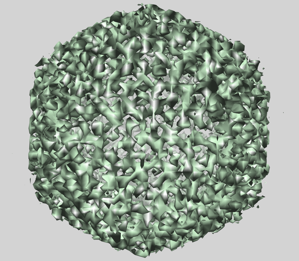 EMD-1575.png EMD-1575.png | 701.3 KB | ||
| Archive directory |  http://ftp.pdbj.org/pub/emdb/structures/EMD-1575ftp://ftp.pdbj.org/pub/emdb/structures/EMD-1575 http://ftp.pdbj.org/pub/emdb/structures/EMD-1575ftp://ftp.pdbj.org/pub/emdb/structures/EMD-1575 | HTTPS FTP |
-Related structure data


-Links
| EMDB pages | EMDB (EBI/PDBe) / EMDataResource |
|---|
-Map
| File | Download / File: emd_1575.map.gz / Format: CCP4 / Size: 166.4 MB / Type: IMAGE STORED AS SIGNED INTEGER (2 BYTES) | ||||||||||||||||||||||||||||||||||||||||||||||||||||||||||||||||||||
|---|---|---|---|---|---|---|---|---|---|---|---|---|---|---|---|---|---|---|---|---|---|---|---|---|---|---|---|---|---|---|---|---|---|---|---|---|---|---|---|---|---|---|---|---|---|---|---|---|---|---|---|---|---|---|---|---|---|---|---|---|---|---|---|---|---|---|---|---|---|
| Annotation | Adenovirus sy12 with anti-sy12 fab | ||||||||||||||||||||||||||||||||||||||||||||||||||||||||||||||||||||
| Projections & slices | Image control
Images are generated by Spider. | ||||||||||||||||||||||||||||||||||||||||||||||||||||||||||||||||||||
| Voxel size | X=Y=Z: 2.54 Å | ||||||||||||||||||||||||||||||||||||||||||||||||||||||||||||||||||||
| Density |
| ||||||||||||||||||||||||||||||||||||||||||||||||||||||||||||||||||||
| Symmetry | Space group: 1 | ||||||||||||||||||||||||||||||||||||||||||||||||||||||||||||||||||||
| Details | EMDB XML:
CCP4 map header:
| ||||||||||||||||||||||||||||||||||||||||||||||||||||||||||||||||||||
 Z (Sec.)
 Y (Row.)
 X (Col.)


-Supplemental data
- Sample components
-Entire : SY12 modified adenovirus 5 plus rabbit anti SY12 Fab
| Entire | Name: SY12 modified adenovirus 5 plus rabbit anti SY12 Fab |
|---|---|
| Components |
|
-Supramolecule #1000: SY12 modified adenovirus 5 plus rabbit anti SY12 Fab
| Supramolecule | Name: SY12 modified adenovirus 5 plus rabbit anti SY12 Fab / type: sample / ID: 1000 Details: dodecapeptide TAYSSYMKGGKF (abbreviated SY12 ) fused to the C-terminus of pIX and a recombinant Ad5LacZ-pIX-SY12 vector has been constructed anti SY12 IgG have been clived into FAb using ...Details: dodecapeptide TAYSSYMKGGKF (abbreviated SY12 ) fused to the C-terminus of pIX and a recombinant Ad5LacZ-pIX-SY12 vector has been constructed anti SY12 IgG have been clived into FAb using papain and then purified. The complex have been generated using an excess of Fab Number unique components: 1 |
|---|
-Supramolecule #1: Human adenovirus 5
| Supramolecule | Name: Human adenovirus 5 / type: virus / ID: 1 / Name.synonym: adenovirus 5 Details: anti SY12 Fab attached to the C-terminal part of adenovirus 5 protein IX NCBI-ID: 28285 / Sci species name: Human adenovirus 5 / Virus type: VIRION / Virus isolate: SEROTYPE / Virus enveloped: No / Virus empty: No / Syn species name: adenovirus 5 |
|---|---|
| Host (natural) | Organism:  Homo sapiens (human) / synonym: VERTEBRATES Homo sapiens (human) / synonym: VERTEBRATES |
| Molecular weight | Experimental: 150 MDa |
| Virus shell | Shell ID: 1 / Diameter: 1000 Å / T number (triangulation number): 25 |
-Experimental details
-Structure determination
| Method | negative staining, cryo EM |
|---|---|
 Processing Processing | single particle reconstruction |
| Aggregation state | particle |
-Sample preparation
| Concentration | 1. mg/mL |
|---|---|
| Buffer | pH: 7.5 / Details: 20mM NaCl, 10mM Tris-HCL |
| Staining | Type: NEGATIVE / Details: Cryo EM |
| Grid | Details: Quantifoil r2/2 |
| Vitrification | Cryogen name: ETHANE / Instrument: OTHER / Details: Vitrification instrument: Zeiss / Method: Blot for 2 seconds before plunging |
- Electron microscopy
| Microscope | JEOL 2010F |
|---|---|
| Electron beam | Acceleration voltage: 200 kV / Electron source: FIELD EMISSION GUN |
| Electron optics | Illumination mode: OTHER / Imaging mode: BRIGHT FIELDBright-field microscopy / Cs: 1.4 mm / Nominal defocus max: 3.5 µm / Nominal defocus min: 1.0 µm / Nominal magnification: 30000 |
| Sample stage | Specimen holder: Eucentric / Specimen holder model: OTHER |
| Alignment procedure | Legacy - Astigmatism: Objective lens astigmatism was corrected at 100,000 times magnification |
| Image recording | Category: FILM / Film or detector model: KODAK SO-163 FILM / Digitization - Scanner: ZEISS SCAI / Digitization - Sampling interval: 7 µm / Number real images: 11 / Average electron dose: 9 e/Å2 / Bits/pixel: 8 |
-Image processing
| CTF correction | Details: Each particle |
|---|---|
| Final reconstruction | Applied symmetry - Point group: I (icosahedral) / Algorithm: OTHER / Resolution.type: BY AUTHOR / Resolution: 22.0 Å / Resolution method: FSC 0.33 CUT-OFF / Software - Name: pft2 em3dr2 / Number images used: 949 |
| Details | Fab-adenovirus complex |
