 Movie
Movie Controller

[English] 日本語
 Yorodumi
- EMDB-1461: Near-atomic resolution using electron cryomicroscopy and single-p... -
+ Open data

- Basic information
| Entry | Database: EMDB / ID: EMD-1461 | |||||||||
|---|---|---|---|---|---|---|---|---|---|---|
| Title | Near-atomic resolution using electron cryomicroscopy and single-particle reconstruction. | |||||||||
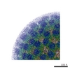
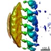
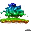
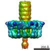
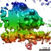
 Map data Map data | Map of rotavirus VP6 protein, after icosahedral averaging and 13-fold non-icosahedral averaging | |||||||||
 Sample Sample |
| |||||||||
| Biological species |  Bovine rotavirus Bovine rotavirus | |||||||||
| Method | single particle reconstruction / cryo EM / negative staining / Resolution: 3.8 Å | |||||||||
 Authors Authors | Zhang X / Settembre E / Xu C / Dormitzer PR / Bellamy R / Harrison SC / Grigorieff N | |||||||||
 Citation Citation | Journal: Proc Natl Acad Sci U S A / Year: 2008 Title: Near-atomic resolution using electron cryomicroscopy and single-particle reconstruction. Authors: Xing Zhang / Ethan Settembre / Chen Xu / Philip R Dormitzer / Richard Bellamy / Stephen C Harrison / Nikolaus Grigorieff /  Abstract: Electron cryomicroscopy (cryo-EM) yields images of macromolecular assemblies and their components, from which 3D structures can be determined, by using an image processing method commonly known as ...Electron cryomicroscopy (cryo-EM) yields images of macromolecular assemblies and their components, from which 3D structures can be determined, by using an image processing method commonly known as "single-particle reconstruction." During the past two decades, this technique has become an important tool for 3D structure determination, but it generally has not been possible to determine atomic models. In principle, individual molecular images contain high-resolution information contaminated by a much higher level of noise. In practice, it has been unclear whether current averaging methods are adequate to extract this information from the background. We present here a reconstruction, obtained by using recently developed image processing methods, of the rotavirus inner capsid particle ("double-layer particle" or DLP) at a resolution suitable for interpretation by an atomic model. The result establishes single-particle reconstruction as a high-resolution technique. We show by direct comparison that the cryo-EM reconstruction of viral protein 6 (VP6) of the rotavirus DLP is similar in clarity to a 3.8-A resolution map obtained from x-ray crystallography. At this resolution, most of the amino acid side chains produce recognizable density. The icosahedral symmetry of the particle was an important factor in achieving this resolution in the cryo-EM analysis, but as the size of recordable datasets increases, single-particle reconstruction also is likely to yield structures at comparable resolution from samples of much lower symmetry. This potential has broad implications for structural cell biology. | |||||||||
| History |
|
- Structure visualization
| Movie |
 Movie viewer Movie viewer |
|---|---|
| Structure viewer | EM map: SurfViewMolmilJmol/JSmol |
| Supplemental images |

- Downloads & links
-EMDB archive
| Map data | emd_1461.map.gz | 1.6 MB | EMDB map data format | |
|---|---|---|---|---|
| Header (meta data) | emd-1461-v30.xmlemd-1461.xml | 9.7 KB 9.7 KB | Display Display | EMDB header |
| Images |  1461.gif 1461.gif | 34.9 KB | ||
| Archive directory |  http://ftp.pdbj.org/pub/emdb/structures/EMD-1461ftp://ftp.pdbj.org/pub/emdb/structures/EMD-1461 http://ftp.pdbj.org/pub/emdb/structures/EMD-1461ftp://ftp.pdbj.org/pub/emdb/structures/EMD-1461 | HTTPS FTP |
-Validation report
| Summary document | emd_1461_validation.pdf.gz | 229.5 KB | Display | EMDB validaton report |
|---|---|---|---|---|
| Full document | emd_1461_full_validation.pdf.gz | 228.5 KB | Display | |
| Data in XML | emd_1461_validation.xml.gz | 4.4 KB | Display | |
| Arichive directory | https://ftp.pdbj.org/pub/emdb/validation_reports/EMD-1461ftp://ftp.pdbj.org/pub/emdb/validation_reports/EMD-1461 | HTTPS FTP |
-Related structure data

-Links
| EMDB pages | EMDB (EBI/PDBe) / EMDataResource |
|---|
-Map
| File | Download / File: emd_1461.map.gz / Format: CCP4 / Size: 6.5 MB / Type: IMAGE STORED AS FLOATING POINT NUMBER (4 BYTES) | ||||||||||||||||||||||||||||||||||||||||||||||||||||||||||||||||||||
|---|---|---|---|---|---|---|---|---|---|---|---|---|---|---|---|---|---|---|---|---|---|---|---|---|---|---|---|---|---|---|---|---|---|---|---|---|---|---|---|---|---|---|---|---|---|---|---|---|---|---|---|---|---|---|---|---|---|---|---|---|---|---|---|---|---|---|---|---|---|
| Annotation | Map of rotavirus VP6 protein, after icosahedral averaging and 13-fold non-icosahedral averaging | ||||||||||||||||||||||||||||||||||||||||||||||||||||||||||||||||||||
| Voxel size | X=Y=Z: 1.23 Å | ||||||||||||||||||||||||||||||||||||||||||||||||||||||||||||||||||||
| Density |
| ||||||||||||||||||||||||||||||||||||||||||||||||||||||||||||||||||||
| Symmetry | Space group: 1 | ||||||||||||||||||||||||||||||||||||||||||||||||||||||||||||||||||||
| Details | EMDB XML:
CCP4 map header:
| ||||||||||||||||||||||||||||||||||||||||||||||||||||||||||||||||||||
-Supplemental data
- Sample components
-Entire : Rotavirus VP6 protein
| Entire | Name: Rotavirus VP6 protein |
|---|---|
| Components |
|
-Supramolecule #1000: Rotavirus VP6 protein
| Supramolecule | Name: Rotavirus VP6 protein / type: sample / ID: 1000 Details: VP6 is one component of rotavirus DLP, which has been deposited separately (accession number EMD-1460) Oligomeric state: 780 molecules of VP6 form a DLP particle with 12 molecules of VP1, 120 molecules of VP2, 12 molecules of VP3 and 11 dsRNA molecules Number unique components: 1 |
|---|---|
| Molecular weight | Theoretical: 41 KDa |
-Macromolecule #1: VP6
| Macromolecule | Name: VP6 / type: protein_or_peptide / ID: 1 / Name.synonym: Virus protein 6 / Recombinant expression: No |
|---|---|
| Source (natural) | Organism: Bovine rotavirus |
| Molecular weight | Experimental: 41 KDa |
-Experimental details
-Structure determination
| Method | negative staining, cryo EM |
|---|---|
 Processing Processing | single particle reconstruction |
| Aggregation state | particle |
-Sample preparation
| Concentration | 5 mg/mL |
|---|---|
| Buffer | pH: 7.4 |
| Staining | Type: NEGATIVE / Details: Ice |
| Grid | Details: Lacy carbon and C-flat |
| Vitrification | Cryogen name: ETHANE / Chamber humidity: 30 % / Instrument: HOMEMADE PLUNGER Details: Vitrification instrument: Home-made. Vitrification carried out in air at room temperature Method: Blot for 3 seconds before plunging |
- Electron microscopy
| Microscope | FEI TECNAI F30 |
|---|---|
| Temperature | Average: 90 K |
| Alignment procedure | Legacy - Astigmatism: Objective lens astigmatism was corrected Legacy - Electron beam tilt params: 0 |
| Date | Jun 1, 2007 |
| Image recording | Category: FILM / Film or detector model: KODAK SO-163 FILM / Digitization - Scanner: ZEISS SCAI / Digitization - Sampling interval: 7 µm / Number real images: 386 / Average electron dose: 15 e/Å2 / Od range: 1 / Bits/pixel: 8 |
| Tilt angle min | 0 |
| Tilt angle max | 0 |
| Electron beam | Acceleration voltage: 300 kV / Electron source:  FIELD EMISSION GUN FIELD EMISSION GUN |
| Electron optics | Calibrated magnification: 56540 / Illumination mode: FLOOD BEAM / Imaging mode: BRIGHT FIELD / Cs: 2 mm / Nominal defocus max: 3.5 µm / Nominal defocus min: 1.1 µm / Nominal magnification: 59000 |
| Sample stage | Specimen holder: Eucentric, side-entry / Specimen holder model: GATAN LIQUID NITROGEN |
| Experimental equipment |  Model: Tecnai F30 / Image courtesy: FEI Company |
-Image processing
| Details | Particles were selected using the computer program SIGNATURE |
|---|---|
| CTF correction | Details: Each particle |
| Final reconstruction | Applied symmetry - Point group: I (icosahedral) / Algorithm: OTHER / Resolution.type: BY AUTHOR / Resolution: 3.8 Å / Resolution method: OTHER / Software - Name: FREALIGN Details: The FREALIGN reconstruction was followed by non-icosahedral averaging using the Uppsala program package for X-ray crystallography Number images used: 8400 |
