Movie
Movie Controller

[English] 日本語
 Yorodumi
- EMDB-1448: Structures of the human pyruvate dehydrogenase complex cores: a h... -
+ Open data

- Basic information
| Entry | Database: EMDB / ID: EMD-1448 | |||||||||
|---|---|---|---|---|---|---|---|---|---|---|
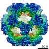
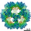
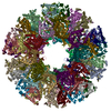
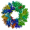
| Title | Structures of the human pyruvate dehydrogenase complex cores: a highly conserved catalytic center with flexible N-terminal domains. | |||||||||
 Map data Map data | This is the half density map for the truncated human dihydrolipoyl acetyltransferase(E2)dodecahedron | |||||||||
 Sample Sample |
| |||||||||
| Function / homology |  Function and homology information Function and homology information dihydrolipoyllysine-residue acetyltransferase / dihydrolipoyllysine-residue acetyltransferase activity / acetyl-CoA biosynthetic process from pyruvate / pyruvate dehydrogenase complex / : / Pyruvate metabolism / Glyoxylate metabolism and glycine degradation / Regulation of pyruvate dehydrogenase (PDH) complex / Signaling by Retinoic Acid / tricarboxylic acid cycle ...dihydrolipoyllysine-residue acetyltransferase / dihydrolipoyllysine-residue acetyltransferase activity / acetyl-CoA biosynthetic process from pyruvate / pyruvate dehydrogenase complex / : / Pyruvate metabolism / Glyoxylate metabolism and glycine degradation / Regulation of pyruvate dehydrogenase (PDH) complex / Signaling by Retinoic Acid / tricarboxylic acid cycle / glucose metabolic process / mitochondrial matrix / intracellular membrane-bounded organelle / mitochondrion / identical protein binding dihydrolipoyllysine-residue acetyltransferase / dihydrolipoyllysine-residue acetyltransferase activity / acetyl-CoA biosynthetic process from pyruvate / pyruvate dehydrogenase complex / : / Pyruvate metabolism / Glyoxylate metabolism and glycine degradation / Regulation of pyruvate dehydrogenase (PDH) complex / Signaling by Retinoic Acid / tricarboxylic acid cycle ...dihydrolipoyllysine-residue acetyltransferase / dihydrolipoyllysine-residue acetyltransferase activity / acetyl-CoA biosynthetic process from pyruvate / pyruvate dehydrogenase complex / : / Pyruvate metabolism / Glyoxylate metabolism and glycine degradation / Regulation of pyruvate dehydrogenase (PDH) complex / Signaling by Retinoic Acid / tricarboxylic acid cycle / glucose metabolic process / mitochondrial matrix / intracellular membrane-bounded organelle / mitochondrion / identical protein bindingSimilarity search - Function | |||||||||
| Biological species |  Homo sapiens (human) Homo sapiens (human) | |||||||||
| Method | single particle reconstruction / cryo EM / Resolution: 8.8 Å | |||||||||
 Authors Authors | Yu X / Hiromasa Y / Tsen H / Stoops JK / Roche TE / Zhou ZH | |||||||||
 Citation Citation | Journal: Structure / Year: 2008 Title: Structures of the human pyruvate dehydrogenase complex cores: a highly conserved catalytic center with flexible N-terminal domains. Authors: Xuekui Yu / Yasuaki Hiromasa / Hua Tsen / James K Stoops / Thomas E Roche / Z Hong Zhou /  Abstract: Dihydrolipoyl acetyltransferase (E2) is the central component of pyruvate dehydrogenase complex (PDC), which converts pyruvate to acetyl-CoA. Structural comparison by cryo-electron microscopy (cryo- ...Dihydrolipoyl acetyltransferase (E2) is the central component of pyruvate dehydrogenase complex (PDC), which converts pyruvate to acetyl-CoA. Structural comparison by cryo-electron microscopy (cryo-EM) of the human full-length and truncated E2 (tE2) cores revealed flexible linkers emanating from the edges of trimers of the internal catalytic domains. Using the secondary structure constraints revealed in our 8 A cryo-EM reconstruction and the prokaryotic tE2 atomic structure as a template, we derived a pseudo atomic model of human tE2. The active sites are conserved between prokaryotic tE2 and human tE2. However, marked structural differences are apparent in the hairpin domain and in the N-terminal helix connected to the flexible linker. These permutations away from the catalytic center likely impart structures needed to integrate a second component into the inner core and provide a sturdy base for the linker that holds the pyruvate dehydrogenase for access by the E2-bound regulatory kinase/phosphatase components in humans. | |||||||||
| History |
|
- Structure visualization
| Movie |
Movie viewer |
|---|---|
| Structure viewer | EM map: SurfViewMolmilJmol/JSmol |
| Supplemental images |

- Downloads & links
-EMDB archive
| Map data | emd_1448.map.gz | 29.5 MB | EMDB map data format | |
|---|---|---|---|---|
| Header (meta data) | emd-1448-v30.xmlemd-1448.xml | 9.9 KB 9.9 KB | Display Display | EMDB header |
| Images | 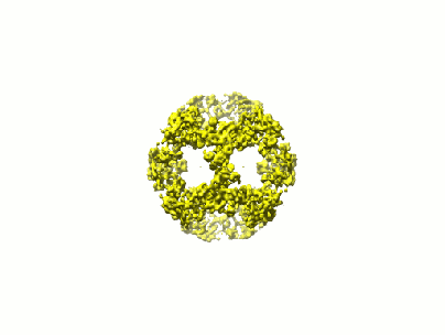 1448.gif 1448.gif | 16.1 KB | ||
| Archive directory |  http://ftp.pdbj.org/pub/emdb/structures/EMD-1448ftp://ftp.pdbj.org/pub/emdb/structures/EMD-1448 http://ftp.pdbj.org/pub/emdb/structures/EMD-1448ftp://ftp.pdbj.org/pub/emdb/structures/EMD-1448 | HTTPS FTP |
-Related structure data
| Related structure data | 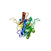 3b8kMC M: atomic model generated by this map C: citing same article ( |
|---|---|
| Similar structure data |


-Links
| EMDB pages | EMDB (EBI/PDBe) / EMDataResource |
|---|---|
| Related items in Molecule of the Month |


-Map
| File | Download / File: emd_1448.map.gz / Format: CCP4 / Size: 55.5 MB / Type: IMAGE STORED AS FLOATING POINT NUMBER (4 BYTES) | ||||||||||||||||||||||||||||||||||||||||||||||||||||||||||||||||||||
|---|---|---|---|---|---|---|---|---|---|---|---|---|---|---|---|---|---|---|---|---|---|---|---|---|---|---|---|---|---|---|---|---|---|---|---|---|---|---|---|---|---|---|---|---|---|---|---|---|---|---|---|---|---|---|---|---|---|---|---|---|---|---|---|---|---|---|---|---|---|
| Annotation | This is the half density map for the truncated human dihydrolipoyl acetyltransferase(E2)dodecahedron | ||||||||||||||||||||||||||||||||||||||||||||||||||||||||||||||||||||
| Voxel size | X=Y=Z: 1.09 Å | ||||||||||||||||||||||||||||||||||||||||||||||||||||||||||||||||||||
| Density |
| ||||||||||||||||||||||||||||||||||||||||||||||||||||||||||||||||||||
| Symmetry | Space group: 1 | ||||||||||||||||||||||||||||||||||||||||||||||||||||||||||||||||||||
| Details | EMDB XML:
CCP4 map header:
| ||||||||||||||||||||||||||||||||||||||||||||||||||||||||||||||||||||
-Supplemental data
- Sample components
-Entire : the truncated human dihydrolipoyl acetyltransferase
| Entire | Name: the truncated human dihydrolipoyl acetyltransferase |
|---|---|
| Components |
|
-Supramolecule #1000: the truncated human dihydrolipoyl acetyltransferase
| Supramolecule | Name: the truncated human dihydrolipoyl acetyltransferase / type: sample / ID: 1000 / Oligomeric state: dodecahedrial assembly of tE2 / Number unique components: 1 |
|---|---|
| Molecular weight | Theoretical: 1.6 MDa |
-Macromolecule #1: truncated human dihydrolipoyl acetyltransferase
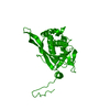
| Macromolecule | Name: truncated human dihydrolipoyl acetyltransferase / type: protein_or_peptide / ID: 1 / Name.synonym: tE2 Details: Human tE2 was prepared from scE2, which contains a PreScission site in the third linker region. Treatment of scE2 with the PreScission protease (Amersham Biosciences) removed the N-terminal 319 amino acids. Number of copies: 60 / Oligomeric state: Dodecahedron / Recombinant expression: Yes |
|---|---|
| Source (natural) | Organism: Homo sapiens (human) / synonym: Human |
| Molecular weight | Experimental: 1.6 MDa / Theoretical: 1.6 MDa |
| Recombinant expression | Organism:  Escherichia coli (E. coli) Escherichia coli (E. coli) |
-Experimental details
-Structure determination
| Method | cryo EM |
|---|---|
 Processing Processing | single particle reconstruction |
| Aggregation state | particle |
-Sample preparation
| Concentration | 0.2 mg/mL |
|---|---|
| Buffer | pH: 7.2 / Details: PBS |
| Grid | Details: 200 mesh holey carbon grid |
| Vitrification | Cryogen name: ETHANE / Instrument: HOMEMADE PLUNGER / Details: Vitrification instrument: lab-made plunger / Method: Blot for 1 second before plunging |
- Electron microscopy
| Microscope | JEOL 2010F |
|---|---|
| Electron beam | Acceleration voltage: 200 kV / Electron source: FIELD EMISSION GUN |
| Electron optics | Calibrated magnification: 69250 / Illumination mode: FLOOD BEAM / Imaging mode: BRIGHT FIELDBright-field microscopy / Cs: 1.0 mm / Nominal defocus max: 2.1 µm / Nominal defocus min: 0.6 µm / Nominal magnification: 69250 |
| Sample stage | Specimen holder: Eucentric / Specimen holder model: GATAN LIQUID NITROGEN |
| Temperature | Min: 100 K / Max: 100 K / Average: 100 K |
| Date | Oct 10, 2003 |
| Image recording | Category: CCD / Film or detector model: GENERIC GATAN / Average electron dose: 12 e/Å2 |
-Image processing
| CTF correction | Details: each image |
|---|---|
| Final reconstruction | Applied symmetry - Point group: I (icosahedral) / Algorithm: OTHER / Resolution.type: BY AUTHOR / Resolution: 8.8 Å / Resolution method: FSC 0.5 CUT-OFF / Software - Name: IMIRS / Number images used: 2432 |