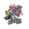
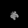
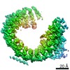
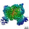
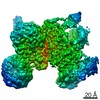
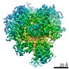
Journal: Science / Year: 2022 Title: Structural basis of branch site recognition by the human spliceosome. Authors: Jonas Tholen / Michal Razew / Felix Weis / Wojciech P Galej / Abstract: Recognition of the intron branch site (BS) by the U2 small nuclear ribonucleoprotein (snRNP) is a critical event during spliceosome assembly. In mammals, BS sequences are poorly conserved, and ...Recognition of the intron branch site (BS) by the U2 small nuclear ribonucleoprotein (snRNP) is a critical event during spliceosome assembly. In mammals, BS sequences are poorly conserved, and unambiguous intron recognition cannot be achieved solely through a base-pairing mechanism. We isolated human 17 U2 snRNP and reconstituted in vitro its adenosine 5´-triphosphate (ATP)–dependent remodeling and binding to the pre–messenger RNA substrate. We determined a series of high-resolution (2.0 to 2.2 angstrom) structures providing snapshots of the BS selection process. The substrate-bound U2 snRNP shows that SF3B6 stabilizes the BS:U2 snRNA duplex, which could aid binding of introns with poor sequence complementarity. ATP-dependent remodeling uncoupled from substrate binding captures U2 snRNA in a conformation that competes with BS recognition, providing a selection mechanism based on branch helix stability.
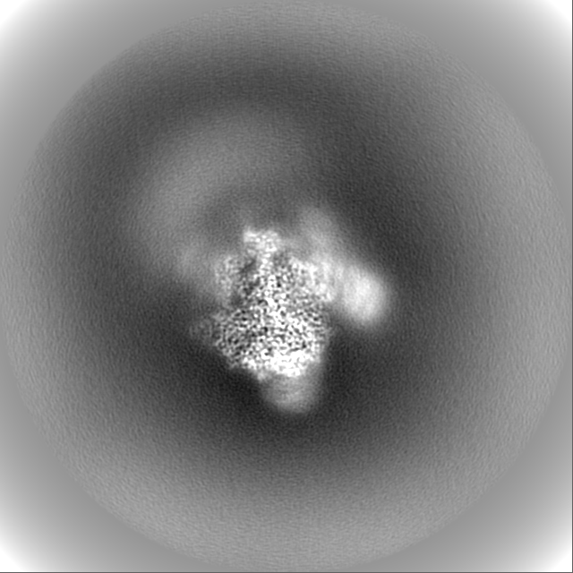
EMPIAR-11000 (Title: Cryo-EM structure of human U2 snRNP after ATP-dependent remodeling Data size: 1.5 TB Data #1: Unaligned multi-frame micrographs of the U2 snRNP after ATP-dependent remodeling [micrographs - multiframe] Data #2: Final polished particles of the U2 snRNP after ATP-dependent remodeling [picked particles - single frame - processed])
Protein or peptide: PHD finger-like domain-containing protein 5A
Ligand: ZINC ION
Ligand: water
+
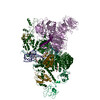
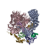
Supramolecule #1: Remodelled U2 snRNP
Supramolecule
Name: Remodelled U2 snRNP / type: complex / ID: 1 / Parent: 0 / Macromolecule list: #1-#8 Details: Remodelled U2 snRNP obtained after incubation of 17S U2 snRNP with ATP. 17S U2 snRNP was isolated from nuclear extract of HEK293F cells by affinity chromatography.
Source (natural)
Organism: Homo sapiens (human)
Molecular weight
Theoretical: 1.08 MDa
+
Macromolecule #1: Splicing factor 3A subunit 2
Macromolecule
Name: Splicing factor 3A subunit 2 / type: protein_or_peptide / ID: 1 / Number of copies: 1 / Enantiomer: LEVO
Film or detector model: GATAN K3 BIOQUANTUM (6k x 4k) / Digitization - Dimensions - Width: 5760 pixel / Digitization - Dimensions - Height: 4092 pixel / Number grids imaged: 2 / Number real images: 15531 / Average exposure time: 1.0 sec. / Average electron dose: 53.45 e/Å2
In the structure databanks used in Yorodumi, some data are registered as the other names, "COVID-19 virus" and "2019-nCoV". Here are the details of the virus and the list of structure data.
Jan 31, 2019. EMDB accession codes are about to change! (news from PDBe EMDB page)
EMDB accession codes are about to change! (news from PDBe EMDB page)
The allocation of 4 digits for EMDB accession codes will soon come to an end. Whilst these codes will remain in use, new EMDB accession codes will include an additional digit and will expand incrementally as the available range of codes is exhausted. The current 4-digit format prefixed with “EMD-” (i.e. EMD-XXXX) will advance to a 5-digit format (i.e. EMD-XXXXX), and so on. It is currently estimated that the 4-digit codes will be depleted around Spring 2019, at which point the 5-digit format will come into force.
The EM Navigator/Yorodumi systems omit the EMD- prefix.
Related info.:Q: What is EMD? / ID/Accession-code notation in Yorodumi/EM Navigator
Yorodumi is a browser for structure data from EMDB, PDB, SASBDB, etc.
This page is also the successor to EM Navigator detail page, and also detail information page/front-end page for Omokage search.
The word "yorodu" (or yorozu) is an old Japanese word meaning "ten thousand". "mi" (miru) is to see.
Related info.:EMDB / PDB / SASBDB / Comparison of 3 databanks / Yorodumi Search / Aug 31, 2016. New EM Navigator & Yorodumi / Yorodumi Papers / Jmol/JSmol / Function and homology information / Changes in new EM Navigator and Yorodumi
 Movie
Movie Controller


 Open data
 Basic information
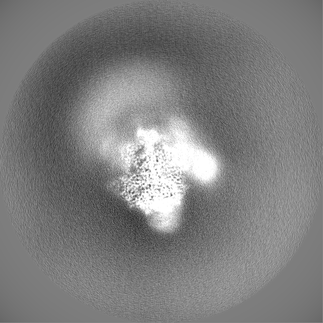
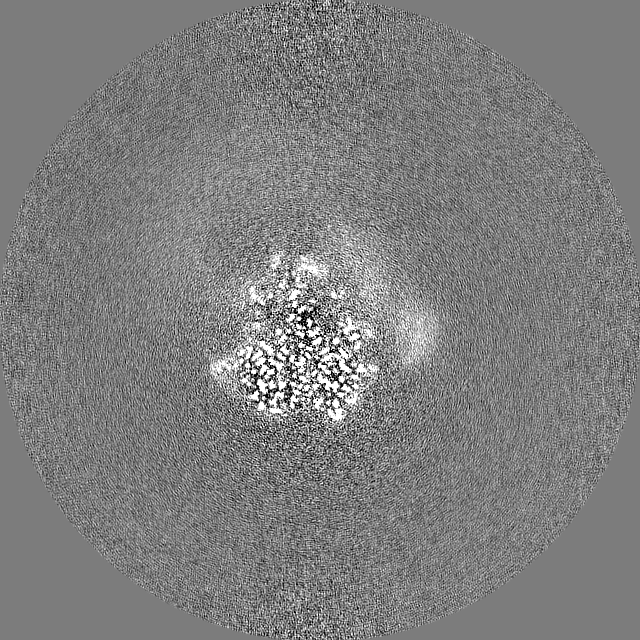
 Map data
 Sample
 Function and homology information
 splicing factor binding / U12-type spliceosomal complex / : /
 Authors
 Citation

 Structure visualization
 Downloads & links
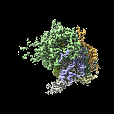 emd_13812.png
 http://ftp.pdbj.org/pub/emdb/structures/EMD-13812


 Z (Sec.)
 Y (Row.)
 X (Col.)


 Sample components
 Processing
 Electron microscopy