Growth arrest and DNA damage-inducible proteins-interacting protein 1 domain superfamily / 39S ribosomal protein L46, mitochondrial / Ribosomal protein L47, mitochondrial / MRP-L47 superfamily, mitochondrial / 39S ribosomal protein L43/54S ribosomal protein L51 / Mitochondrial 39-S ribosomal protein L47 (MRP-L47) / Ribosomal protein L25, beta domain / Ribosomal protein L25, C-terminal / Ribosomal protein TL5, C-terminal domain / Ribosomal protein/NADH dehydrogenase domain ...Growth arrest and DNA damage-inducible proteins-interacting protein 1 domain superfamily / 39S ribosomal protein L46, mitochondrial / Ribosomal protein L47, mitochondrial / MRP-L47 superfamily, mitochondrial / 39S ribosomal protein L43/54S ribosomal protein L51 / Mitochondrial 39-S ribosomal protein L47 (MRP-L47) / Ribosomal protein L25, beta domain / Ribosomal protein L25, C-terminal / Ribosomal protein TL5, C-terminal domain / Ribosomal protein/NADH dehydrogenase domain / Mitochondrial ribosomal protein L51 / S25 / CI-B8 domain / Mitochondrial ribosomal protein L51 / S25 / CI-B8 domain / NUDIX hydrolase-like domain superfamily / Ribosomal protein L6, conserved site / Ribosomal protein L6 signature 1. / Ribosomal protein L17 signature. / Ribosomal protein L9, C-terminal domain superfamily / Ribosomal protein L28/L24 superfamily / Ribosomal protein L25/Gln-tRNA synthetase, anti-codon-binding domain superfamily / Ribosomal protein L9, N-terminal domain superfamily / Ribosomal protein L9 / Ribosomal protein L9, N-terminal / Ribosomal protein L9, N-terminal domain / Ribosomal protein L6, bacterial-type / Ribosomal protein L9/RNase H1, N-terminal / Ribosomal protein L20 signature. / Ribosomal protein L27, conserved site / Ribosomal protein L27 signature. / Ribosomal protein L22, bacterial/chloroplast-type / Ribosomal protein L2, bacterial/organellar-type / Ribosomal L28 family / Ribosomal protein L33 / Ribosomal protein L33 / Ribosomal protein L28/L24 / Ribosomal protein L33 superfamily / Ribosomal protein L30, bacterial-type / Ribosomal protein L16 / L28p-like / Ribosomal protein L20 / Ribosomal protein L20 / Ribosomal protein L20, C-terminal / Ribosomal protein L21 / Ribosomal protein L27 / Ribosomal L27 protein / Ribosomal protein L19 / Ribosomal protein L19 superfamily / Ribosomal protein L19 / Ribosomal proteins 50S L24/mitochondrial 39S L24 / Ribosomal protein L17 / Ribosomal protein L17 superfamily / Ribosomal protein L17 / Ribosomal protein L21-like / L21-like superfamily / Ribosomal prokaryotic L21 protein / Ribosomal L32p protein family / Ribosomal protein L24 / Ribosomal protein L32p / Ribosomal protein L34 / Ribosomal protein L34 / Ribosomal protein L13, bacterial-type / Ribosomal protein L3, bacterial/organelle-type / Ribosomal protein L15, bacterial-type / 50S ribosomal protein uL4 / Ribosomal protein L5 domain superfamily / Ribosomal protein L2, conserved site / Ribosomal protein L2 signature. / Ribosomal protein L10e/L16 / Ribosomal protein L10e/L16 superfamily / Ribosomal protein L6, alpha-beta domain / Ribosomal protein L6 / Ribosomal protein L6, alpha-beta domain superfamily / Ribosomal protein L16p/L10e / Ribosomal protein L6 / Ribosomal protein L2, domain 3 / Ribosomal protein L14P, conserved site / Ribosomal protein L24/L26, conserved site / KOW (Kyprides, Ouzounis, Woese) motif. / Ribosomal Proteins L2, C-terminal domain / Ribosomal protein L2, C-terminal / Ribosomal Proteins L2, C-terminal domain / Ribosomal Proteins L2, RNA binding domain / Ribosomal Proteins L2, RNA binding domain / Ribosomal protein L2 / Ribosomal protein L14 signature. / Ribosomal protein L15 / Ribosomal Proteins L2, RNA binding domain / Ribosomal protein L30, ferredoxin-like fold domain / Ribosomal protein L25/L23 / Ribosomal protein L30, ferredoxin-like fold domain superfamily / Ribosomal protein L14p/L23e / Ribosomal protein L30p/L7e / Ribosomal protein L23 / Ribosomal protein L14P / Ribosomal protein L14 superfamily / Ribosomal proteins 50S-L15, 50S-L18e, 60S-L27A / Ribosomal protein L26/L24, KOW domain / Ribosomal protein L24 signature. / Ribosomal protein L14p/L23e / Ribosomal protein L22/L17 / Ribosomal protein L22/L17 superfamily Similarity search - Domain/homology
Uncharacterized protein / Uncharacterized protein / Uncharacterized protein / Uncharacterized protein / Uncharacterized protein / Ribosomal_L18e/L15P domain-containing protein / Uncharacterized protein / Uncharacterized protein / Uncharacterized protein / KOW domain-containing protein ...Uncharacterized protein / Uncharacterized protein / Uncharacterized protein / Uncharacterized protein / Uncharacterized protein / Ribosomal_L18e/L15P domain-containing protein / Uncharacterized protein / Uncharacterized protein / Uncharacterized protein / KOW domain-containing protein / Uncharacterized protein / Ribosomal_L2_C domain-containing protein / Ribosomal_L16 domain-containing protein / Uncharacterized protein / 50S ribosomal protein L9, chloroplastic / Uncharacterized protein / Ribosomal_L30 domain-containing protein / Uncharacterized protein / Uncharacterized protein / Uncharacterized protein / Uncharacterized protein / Uncharacterized protein / Ribosomal_TL5_C domain-containing protein / Uncharacterized protein / Uncharacterized protein / 50S ribosomal protein L20 / Predicted protein / Predicted protein / Predicted protein / Mitochondrial ribosomal protein L19 / Uncharacterized protein / Mitochondrial ribosomal protein L17 / Predicted protein / Predicted protein / Predicted protein / Plastid ribosomal protein L6 / Predicted protein / Mitochondrial ribosomal protein L23 / Mitochondrial ribosomal protein L13 / Mitochondrial ribosomal protein L33 / Predicted protein / Mitochondrial ribosomal protein L17 Similarity search - Component
Biological species
Chlamydomonas reinhardtii (plant)
Method
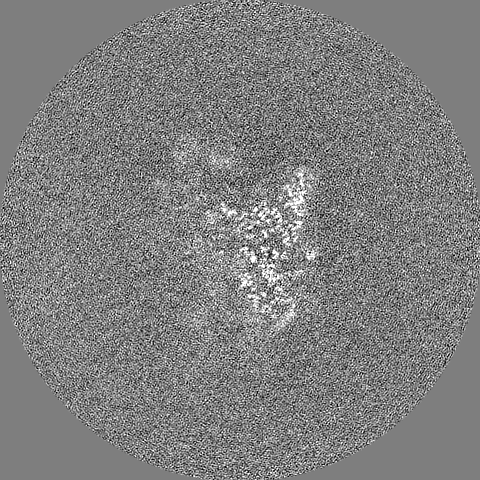
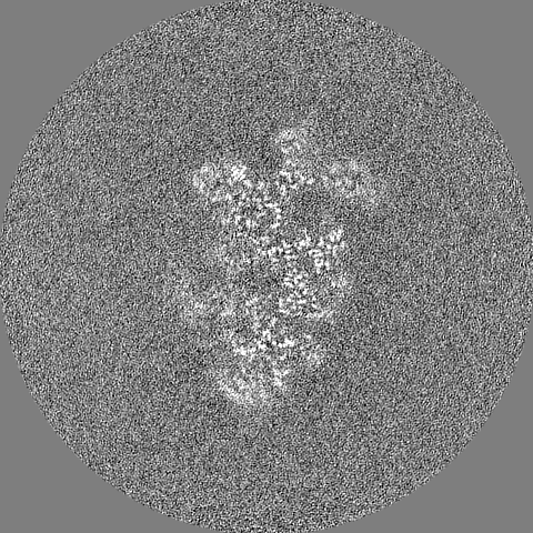
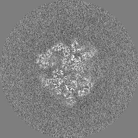
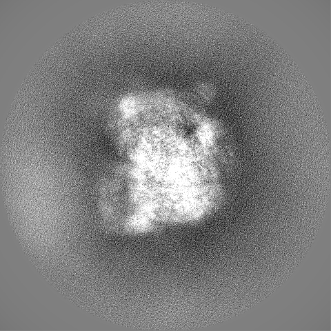
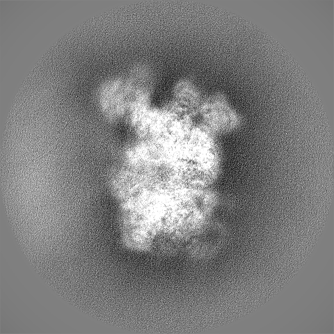
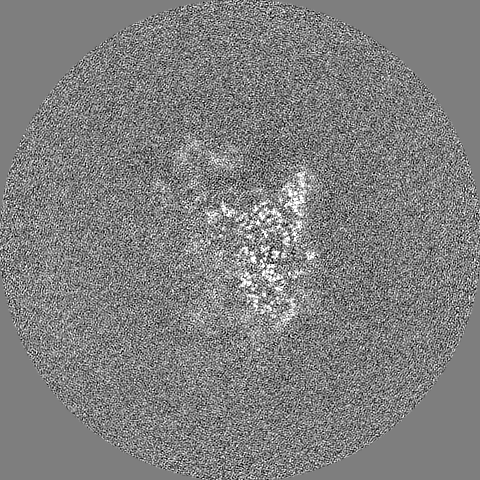
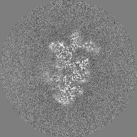
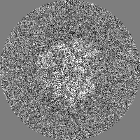
single particle reconstruction / cryo EM / Resolution: 3.0 Å
Journal: Nat Commun / Year: 2021 Title: How to build a ribosome from RNA fragments in Chlamydomonas mitochondria. Authors: Florent Waltz / Thalia Salinas-Giegé / Robert Englmeier / Herrade Meichel / Heddy Soufari / Lauriane Kuhn / Stefan Pfeffer / Friedrich Förster / Benjamin D Engel / Philippe Giegé / ...Authors: Florent Waltz / Thalia Salinas-Giegé / Robert Englmeier / Herrade Meichel / Heddy Soufari / Lauriane Kuhn / Stefan Pfeffer / Friedrich Förster / Benjamin D Engel / Philippe Giegé / Laurence Drouard / Yaser Hashem / Abstract: Mitochondria are the powerhouse of eukaryotic cells. They possess their own gene expression machineries where highly divergent and specialized ribosomes, named hereafter mitoribosomes, translate the ...Mitochondria are the powerhouse of eukaryotic cells. They possess their own gene expression machineries where highly divergent and specialized ribosomes, named hereafter mitoribosomes, translate the few essential messenger RNAs still encoded by mitochondrial genomes. Here, we present a biochemical and structural characterization of the mitoribosome in the model green alga Chlamydomonas reinhardtii, as well as a functional study of some of its specific components. Single particle cryo-electron microscopy resolves how the Chlamydomonas mitoribosome is assembled from 13 rRNA fragments encoded by separate non-contiguous gene pieces. Additional proteins, mainly OPR, PPR and mTERF helical repeat proteins, are found in Chlamydomonas mitoribosome, revealing the structure of an OPR protein in complex with its RNA binding partner. Targeted amiRNA silencing indicates that these ribosomal proteins are required for mitoribosome integrity. Finally, we use cryo-electron tomography to show that Chlamydomonas mitoribosomes are attached to the inner mitochondrial membrane via two contact points mediated by Chlamydomonas-specific proteins. Our study expands our understanding of mitoribosome diversity and the various strategies these specialized molecular machines adopt for membrane tethering.
Macromolecule #10: Mitochondrial ribosomal protein L17,bL17m
Macromolecule
Name: Mitochondrial ribosomal protein L17,bL17m / type: protein_or_peptide / ID: 10 Details: The protein is a mixture of A0A2K3DXS2 and A8JH49,The protein is a mixture of A0A2K3DXS2 and A8JH49 Number of copies: 1 / Enantiomer: LEVO
Name: bL27m / type: protein_or_peptide / ID: 18 Details: N-ter is extend compared to A0A2K3E880,N-ter is extend compared to A0A2K3E880,N-ter is extend compared to A0A2K3E880,N-ter is extend compared to A0A2K3E880 Number of copies: 1 / Enantiomer: LEVO
In the structure databanks used in Yorodumi, some data are registered as the other names, "COVID-19 virus" and "2019-nCoV". Here are the details of the virus and the list of structure data.
Jan 31, 2019. EMDB accession codes are about to change! (news from PDBe EMDB page)
EMDB accession codes are about to change! (news from PDBe EMDB page)
The allocation of 4 digits for EMDB accession codes will soon come to an end. Whilst these codes will remain in use, new EMDB accession codes will include an additional digit and will expand incrementally as the available range of codes is exhausted. The current 4-digit format prefixed with “EMD-” (i.e. EMD-XXXX) will advance to a 5-digit format (i.e. EMD-XXXXX), and so on. It is currently estimated that the 4-digit codes will be depleted around Spring 2019, at which point the 5-digit format will come into force.
The EM Navigator/Yorodumi systems omit the EMD- prefix.
Related info.:Q: What is EMD? / ID/Accession-code notation in Yorodumi/EM Navigator
Yorodumi is a browser for structure data from EMDB, PDB, SASBDB, etc.
This page is also the successor to EM Navigator detail page, and also detail information page/front-end page for Omokage search.
The word "yorodu" (or yorozu) is an old Japanese word meaning "ten thousand". "mi" (miru) is to see.
Related info.:EMDB / PDB / SASBDB / Comparison of 3 databanks / Yorodumi Search / Aug 31, 2016. New EM Navigator & Yorodumi / Yorodumi Papers / Jmol/JSmol / Function and homology information / Changes in new EM Navigator and Yorodumi
 Movie
Movie Controller


 Open data
 Basic information
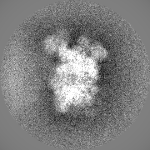
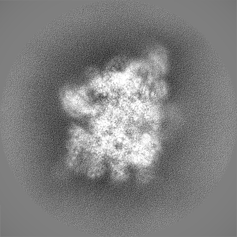
 Map data
 Sample
 Function and homology information
 mitochondrial ribosome / mitochondrial translation /

 Authors
 France, 4 items
  Citation

 Structure visualization
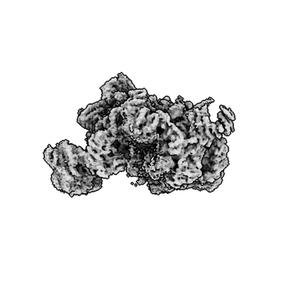
 Downloads & links
 emd_13480.png
 http://ftp.pdbj.org/pub/emdb/structures/EMD-13480
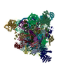


 Z (Sec.)
 Y (Row.)
 X (Col.)


 Sample components
 Processing
 Electron microscopy
