Movie
Movie Controller


+ Open data

- Basic information
| Entry | Database: EMDB / ID: EMD-13071 | |||||||||
|---|---|---|---|---|---|---|---|---|---|---|
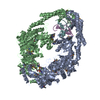
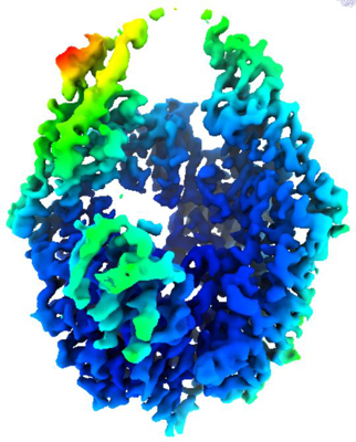
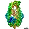
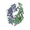
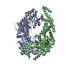
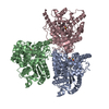
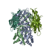
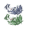
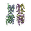
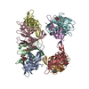
| Title | The structure of MutS bound to two molecules of ADP-Vanadate | |||||||||
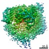
 Map data Map data | ||||||||||
 Sample Sample |
| |||||||||
| Function / homology |  Function and homology information Function and homology informationadenine/cytosine mispair binding /  MutS complex / mismatch repair complex / regulation of DNA recombination / mismatched DNA binding / DNA binding, bending / ATP-dependent DNA damage sensor activity / mismatch repair / ADP binding / damaged DNA binding ...adenine/cytosine mispair binding / MutS complex / mismatch repair complex / regulation of DNA recombination / mismatched DNA binding / DNA binding, bending / ATP-dependent DNA damage sensor activity / mismatch repair / ADP binding / damaged DNA binding / DNA damage response / ATP hydrolysis activity / ATP binding / identical protein binding / cytosol MutS complex / mismatch repair complex / regulation of DNA recombination / mismatched DNA binding / DNA binding, bending / ATP-dependent DNA damage sensor activity / mismatch repair / ADP binding / damaged DNA binding ...adenine/cytosine mispair binding / MutS complex / mismatch repair complex / regulation of DNA recombination / mismatched DNA binding / DNA binding, bending / ATP-dependent DNA damage sensor activity / mismatch repair / ADP binding / damaged DNA binding / DNA damage response / ATP hydrolysis activity / ATP binding / identical protein binding / cytosolSimilarity search - Function | |||||||||
| Biological species |  Escherichia coli (E. coli) / Escherichia coli (strain K12) (bacteria) Escherichia coli (E. coli) / Escherichia coli (strain K12) (bacteria) | |||||||||
| Method | single particle reconstruction / cryo EM / Resolution: 3.8 Å | |||||||||
 Authors Authors | Lamers MH / Borsellini A / Friedhoff P / Kunetsky V | |||||||||
| Funding support | European Union, 1 items
| |||||||||
 Citation Citation | Journal: Nat Struct Mol Biol / Year: 2022 Title: Cryogenic electron microscopy structures reveal how ATP and DNA binding in MutS coordinates sequential steps of DNA mismatch repair. Authors: Alessandro Borsellini / Vladislav Kunetsky / Peter Friedhoff / Meindert H Lamers /   Abstract: DNA mismatch repair detects and corrects mismatches introduced during DNA replication. The protein MutS scans for mismatches and coordinates the repair cascade. During this process, MutS undergoes ...DNA mismatch repair detects and corrects mismatches introduced during DNA replication. The protein MutS scans for mismatches and coordinates the repair cascade. During this process, MutS undergoes multiple conformational changes in response to ATP binding, hydrolysis and release, but how ATP induces the various MutS conformations is incompletely understood. Here we present four cryogenic electron microscopy structures of Escherichia coli MutS at sequential stages of the ATP hydrolysis cycle that reveal how ATP binding and hydrolysis induce closing and opening of the MutS dimer, respectively. Biophysical analysis demonstrates how DNA binding modulates the ATPase cycle by prevention of hydrolysis during scanning and mismatch binding, while preventing ADP release in the sliding clamp state. Nucleotide release is achieved when MutS encounters single-stranded DNA that is produced during removal of the daughter strand. The combination of ATP binding and hydrolysis and its modulation by DNA enables MutS to adopt the different conformations needed to coordinate the sequential steps of the mismatch repair cascade. | |||||||||
| History |
|
- Structure visualization
| Movie |
Movie viewer |
|---|---|
| Structure viewer | EM map: SurfViewMolmilJmol/JSmol |
| Supplemental images |
- Downloads & links
-EMDB archive
| Map data | emd_13071.map.gz | 1.1 MB | EMDB map data format | |
|---|---|---|---|---|
| Header (meta data) | emd-13071-v30.xmlemd-13071.xml | 16.7 KB 16.7 KB | Display Display | EMDB header |
| FSC (resolution estimation) | emd_13071_fsc.xml | 6.9 KB | Display | FSC data file |
| Images |  emd_13071.png emd_13071.png | 171.4 KB | ||
| Archive directory |  http://ftp.pdbj.org/pub/emdb/structures/EMD-13071ftp://ftp.pdbj.org/pub/emdb/structures/EMD-13071 http://ftp.pdbj.org/pub/emdb/structures/EMD-13071ftp://ftp.pdbj.org/pub/emdb/structures/EMD-13071 | HTTPS FTP |
-Related structure data
| Related structure data | 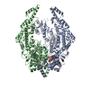 7ou0MC 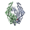 7otoC 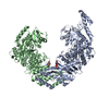 7ou2C 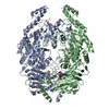 7ou4C M: atomic model generated by this map C: citing same article ( |
|---|---|
| Similar structure data |


-Links
| EMDB pages | EMDB (EBI/PDBe) / EMDataResource |
|---|
-Map
| File | Download / File: emd_13071.map.gz / Format: CCP4 / Size: 3.6 MB / Type: IMAGE STORED AS FLOATING POINT NUMBER (4 BYTES) | ||||||||||||||||||||||||||||||||||||||||||||||||||||||||||||
|---|---|---|---|---|---|---|---|---|---|---|---|---|---|---|---|---|---|---|---|---|---|---|---|---|---|---|---|---|---|---|---|---|---|---|---|---|---|---|---|---|---|---|---|---|---|---|---|---|---|---|---|---|---|---|---|---|---|---|---|---|---|
| Voxel size | X=Y=Z: 1.32 Å | ||||||||||||||||||||||||||||||||||||||||||||||||||||||||||||
| Density |
| ||||||||||||||||||||||||||||||||||||||||||||||||||||||||||||
| Symmetry | Space group: 1 | ||||||||||||||||||||||||||||||||||||||||||||||||||||||||||||
| Details | EMDB XML:
CCP4 map header:
| ||||||||||||||||||||||||||||||||||||||||||||||||||||||||||||
-Supplemental data
- Sample components
-Entire : DNA mismatch repair protein MutS
| Entire | Name: DNA mismatch repair protein MutS |
|---|---|
| Components |
|
-Supramolecule #1: DNA mismatch repair protein MutS
| Supramolecule | Name: DNA mismatch repair protein MutS / type: organelle_or_cellular_component / ID: 1 / Parent: 0 / Macromolecule list: #1 / Details: MutS bound to two molecules of AMPPNP |
|---|---|
| Source (natural) | Organism: Escherichia coli (E. coli) |
| Molecular weight | Theoretical: 190 KDa |
| Recombinant expression | Organism: Escherichia coli 'BL21-Gold(DE3)pLysS AG' (bacteria) |
-Macromolecule #1: DNA mismatch repair protein MutS
| Macromolecule | Name: DNA mismatch repair protein MutS / type: protein_or_peptide / ID: 1 Details: ADP Vanadate ATP hydrolysis transition state analogue. Number of copies: 2 / Enantiomer: LEVO |
|---|---|
| Source (natural) | Organism: Escherichia coli (strain K12) (bacteria) / Strain: K12 |
| Molecular weight | Theoretical: 90.433234 KDa |
| Recombinant expression | Organism: Escherichia coli (E. coli) |
| Sequence | String: HHHHHHMSAI ENFDAHTPMM QQYLRLKAQH PEILLFYRMG DFYELFYDDA KRASQLLDIS LTKRGASAGE PIPMAGIPYH AVENYLAKL VNQGESVAIC EQIGDPATSK GPVERKVVRI VTPGTISDEA LLQERQDNLL AAIWQDSKGF GYATLDISSG R FRLSEPAD ...String: HHHHHHMSAI ENFDAHTPMM QQYLRLKAQH PEILLFYRMG DFYELFYDDA KRASQLLDIS LTKRGASAGE PIPMAGIPYH AVENYLAKL VNQGESVAIC EQIGDPATSK GPVERKVVRI VTPGTISDEA LLQERQDNLL AAIWQDSKGF GYATLDISSG R FRLSEPAD RETMAAELQR TNPAELLYAE DFAEMSLIEG RRGLRRRPLW EFEIDTARQQ LNLQFGTRDL VGFGVENAPR GL CAAGCLL QYAKDTQRTT LPHIRSITME REQDSIIMDA ATRRNLEITQ NLAGGAENTL ASVLDCTVTP MGSRMLKRWL HMP VRDTRV LLERQQTIGA LQDFTAGLQP VLRQVGDLER ILARLALRTA RPRDLARMRH AFQQLPELRA QLETVDSAPV QALR EKMGE FAELRDLLER AIIDTPPVLV RDGGVIASGY NEELDEWRAL ADGATDYLER LEVRERERTG LDTLKVGFNA VHGYY IQIS RGQSHLAPIN YMRRQTLKNA ERYIIPELKE YEDKVLTSKG KALALEKQLY EELFDLLLPH LEALQQSASA LAELDV LVN LAERAYTLNY TCPTFIDKPG IRITEGRHPV VEQVLNEPFI ANPLNLSPQR RMLIITGPNM GGKSTYMRQT ALIALMA YI GSYVPAQKVE IGPIDRIFTR VGAADDLASG RSTFMVEMTE TANILHNATE YSLVLMDEIG RGTSTYDGLS LAWACAEN L ANKIKALTLF ATHYFELTQL PEKMEGVANV HLDALEHGDT IAFMHSVQDG AASKSYGLAV AALAGVPKEV IKRARQKLR ELESIS |
-Macromolecule #2: MAGNESIUM ION
| Macromolecule | Name: MAGNESIUM ION / type: ligand / ID: 2 / Number of copies: 2 / Formula: MG |
|---|---|
| Molecular weight | Theoretical: 24.305 Da |
-Macromolecule #3: ADENOSINE-5'-DIPHOSPHATE
| Macromolecule | Name: ADENOSINE-5'-DIPHOSPHATE / type: ligand / ID: 3 / Number of copies: 2 / Formula: ADP |
|---|---|
| Molecular weight | Theoretical: 427.201 Da |
| Chemical component information |  ChemComp-ADP: |
-Macromolecule #4: VANADATE ION
| Macromolecule | Name: VANADATE ION / type: ligand / ID: 4 / Number of copies: 2 / Formula: VO4 |
|---|---|
| Molecular weight | Theoretical: 114.939 Da |
| Chemical component information |  ChemComp-VN3: |
-Experimental details
-Structure determination
| Method | cryo EM |
|---|---|
 Processing Processing | single particle reconstruction |
| Aggregation state | particle |
-Sample preparation
| Concentration | 0.95 mg/mL | ||||||||||||||||||
|---|---|---|---|---|---|---|---|---|---|---|---|---|---|---|---|---|---|---|---|
| Buffer | pH: 8.5 Component:
| ||||||||||||||||||
| Grid | Model: Quantifoil R1.2/1.3 / Material: COPPER / Mesh: 300 / Support film - Material: CARBON / Support film - topology: HOLEY / Pretreatment - Type: GLOW DISCHARGE / Pretreatment - Atmosphere: AIR / Pretreatment - Pressure: 0.0004 kPa | ||||||||||||||||||
| Vitrification | Cryogen name: ETHANE / Chamber humidity: 76 % / Chamber temperature: 4 K / Instrument: LEICA PLUNGER |
- Electron microscopy
| Microscope | FEI TITAN KRIOS |
|---|---|
| Electron beam | Acceleration voltage: 300 kV / Electron source: FIELD EMISSION GUN |
| Electron optics | Illumination mode: FLOOD BEAM / Imaging mode: BRIGHT FIELDBright-field microscopy / Cs: 2.7 mm / Nominal defocus max: 2.0 µm / Nominal defocus min: 0.5 µm / Nominal magnification: 105000 |
| Specialist optics | Energy filter - Name: GIF Bioquantum / Energy filter - Slit width: 20 eV |
| Sample stage | Specimen holder model: FEI TITAN KRIOS AUTOGRID HOLDER / Cooling holder cryogen: NITROGEN |
| Image recording | Film or detector model: GATAN K2 SUMMIT (4k x 4k) / Detector mode: COUNTING / Digitization - Frames/image: 1-50 / Number real images: 4835 / Average electron dose: 57.0 e/Å2 |
| Experimental equipment |  Model: Titan Krios / Image courtesy: FEI Company |
-Image processing
| Particle selection | Number selected: 496416 |
|---|---|
| CTF correction | Software - Name: Gctf (ver. 1.06) |
| Startup model | Type of model: OTHER Details: density map coming from MutS bound to two molecules of AMPPNP. density map reported together with this one in the same manuscript. |
| Initial angle assignment | Type: MAXIMUM LIKELIHOOD / Software - Name: RELION (ver. 3.1) |
| Final 3D classification | Number classes: 4 / Software - Name: RELION (ver. 3.1) |
| Final angle assignment | Type: MAXIMUM LIKELIHOOD / Software - Name: RELION (ver. 3.1) |
| Final reconstruction | Resolution.type: BY AUTHOR / Resolution: 3.8 Å / Resolution method: FSC 0.143 CUT-OFF / Software - Name: REFMAC / Number images used: 81316 |
| FSC plot (resolution estimation) | 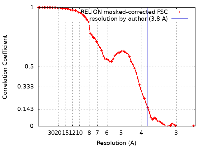 |