 Movie
Movie Controller

[English] 日本語
 Yorodumi
- EMDB-1273: Centrosome polarization delivers secretory granules to the immuno... -
+ Open data

- Basic information
| Entry | Database: EMDB / ID: EMD-1273 | |||||||||
|---|---|---|---|---|---|---|---|---|---|---|
| Title | Centrosome polarization delivers secretory granules to the immunological synapse. | |||||||||
 Map data Map data | Tomogram of the immunological synapse between a cytotoxic T lymphocyte (CTL) and a target cell. The microtubule organization centre (MTOC) is polarized to the cell-cell contact site. The electron-dense lytic granules are transported along the microtubules to the synaptic cleft. Here they probably fuse with the membrane and excrete the cytotoxic proteins that trigger the death of the target cell. (See the masks representing models of the synaptic cleft, microtubules, centriole, lytic granules and Golgi apparatus.) | |||||||||
 Sample Sample |
| |||||||||
| Biological species |   Homo sapiens (human) / Homo sapiens (human) /  Mus musculus (house mouse) Mus musculus (house mouse) | |||||||||
| Method | electron tomography / negative staining / Resolution: 50.0 Å | |||||||||
 Authors Authors | Stinchcombe JC / Majorovits E / Bossi G / Fuller SD / Griffiths GM | |||||||||
 Citation Citation | Journal: Nature / Year: 2006 Title: Centrosome polarization delivers secretory granules to the immunological synapse. Authors: Jane C Stinchcombe / Endre Majorovits / Giovanna Bossi / Stephen Fuller / Gillian M Griffiths /  Abstract: Cytotoxic T lymphocytes (CTLs) destroy virally infected and tumorigenic cells by releasing the contents of specialized secretory lysosomes--termed 'lytic granules'--at the immunological synapse ...Cytotoxic T lymphocytes (CTLs) destroy virally infected and tumorigenic cells by releasing the contents of specialized secretory lysosomes--termed 'lytic granules'--at the immunological synapse formed between the CTL and the target. On contact with the target cell, the microtubule organizing centre of the CTL polarizes towards the target and granules move along microtubules in a minus-end direction towards the polarized microtubule organizing centre. However, the final steps of secretion have remained unclear. Here we show that CTLs do not require actin or plus-end microtubule motors for secretion, but instead the centrosome moves to and contacts the plasma membrane at the central supramolecular activation cluster of the immunological synapse. Actin and IQGAP1 are cleared away from the synapse, and granules are delivered directly to the plasma membrane. These data show that CTLs use a previously unreported mechanism for delivering secretory granules to the immunological synapse, with granule secretion controlled by centrosome delivery to the plasma membrane. | |||||||||
| History |
|
- Structure visualization
| Movie |
 Movie viewer Movie viewer |
|---|---|
| Structure viewer | EM map: SurfViewMolmilJmol/JSmol |
| Supplemental images |

- Downloads & links
-EMDB archive
| Map data | emd_1273.map.gz | 196.6 MB | EMDB map data format | |
|---|---|---|---|---|
| Header (meta data) | emd-1273-v30.xmlemd-1273.xml | 21.9 KB 21.9 KB | Display Display | EMDB header |
| Images | 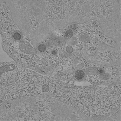 1273.gif 1273.gif | 139.9 KB | ||
| Masks | emd_1273_msk_1.mapemd_1273_msk_2.mapemd_1273_msk_3.mapemd_1273_msk_4.mapemd_1273_msk_5.map | 304 MB 304 MB 304 MB 304 MB 304 MB | Mask map | |
| Others | README_masks.txt | 1.6 KB | ||
| Archive directory |  http://ftp.pdbj.org/pub/emdb/structures/EMD-1273ftp://ftp.pdbj.org/pub/emdb/structures/EMD-1273 http://ftp.pdbj.org/pub/emdb/structures/EMD-1273ftp://ftp.pdbj.org/pub/emdb/structures/EMD-1273 | HTTPS FTP |
-Links
| EMDB pages | EMDB (EBI/PDBe) / EMDataResource |
|---|
-Map
| File | Download / File: emd_1273.map.gz / Format: CCP4 / Size: 296.9 MB / Type: IMAGE STORED AS SIGNED BYTE | ||||||||||||||||||||||||||||||||||||||||||||||||||||||||||||||||||||
|---|---|---|---|---|---|---|---|---|---|---|---|---|---|---|---|---|---|---|---|---|---|---|---|---|---|---|---|---|---|---|---|---|---|---|---|---|---|---|---|---|---|---|---|---|---|---|---|---|---|---|---|---|---|---|---|---|---|---|---|---|---|---|---|---|---|---|---|---|---|
| Annotation | Tomogram of the immunological synapse between a cytotoxic T lymphocyte (CTL) and a target cell. The microtubule organization centre (MTOC) is polarized to the cell-cell contact site. The electron-dense lytic granules are transported along the microtubules to the synaptic cleft. Here they probably fuse with the membrane and excrete the cytotoxic proteins that trigger the death of the target cell. (See the masks representing models of the synaptic cleft, microtubules, centriole, lytic granules and Golgi apparatus.) | ||||||||||||||||||||||||||||||||||||||||||||||||||||||||||||||||||||
| Projections & slices | Image control
Images are generated by Spider. generated in cubic-lattice coordinate | ||||||||||||||||||||||||||||||||||||||||||||||||||||||||||||||||||||
| Voxel size | X=Y=Z: 22.5 Å | ||||||||||||||||||||||||||||||||||||||||||||||||||||||||||||||||||||
| Density |
| ||||||||||||||||||||||||||||||||||||||||||||||||||||||||||||||||||||
| Symmetry | Space group: 1 | ||||||||||||||||||||||||||||||||||||||||||||||||||||||||||||||||||||
| Details | EMDB XML:
CCP4 map header:
| ||||||||||||||||||||||||||||||||||||||||||||||||||||||||||||||||||||
 Z (Sec.)
 Y (Row.)
 X (Col.)


-Supplemental data
-Segmentation: Microtubule (MT) network within the CTL. The MTs...
| Annotation | Microtubule (MT) network within the CTL. The MTs radiate out from the polarized centrosomal area, running both away from the plasma membrane into the cell and along the cell membrane to the periphery of the synapse. Both lytic granules (see mask granules_mask4) and the Golgi apparatus (see mask golgi_mask5) are linked to and transported along the MTs to the synaptic cleft | ||||||||||||
|---|---|---|---|---|---|---|---|---|---|---|---|---|---|
| File | emd_1273_msk_1.map | ||||||||||||
| Projections & Slices |
| ||||||||||||
| Density Histograms |


-Segmentation: One of the two CTL centrioles, sitting close...
| Annotation | One of the two CTL centrioles, sitting close to the synaptic cleft. The two centrioles form part of the centrosome that is polarized to the synaptic cleft. Microtubules run from here into the cell and along the cell membrane to the periphery of the synapse (see mask MTs_mask) | ||||||||||||
|---|---|---|---|---|---|---|---|---|---|---|---|---|---|
| File | emd_1273_msk_2.map | ||||||||||||
| Projections & Slices |
| ||||||||||||
| Density Histograms |


-Segmentation: The lytic granules are the "cytotoxic organelles" of...
| Annotation | The lytic granules are the "cytotoxic organelles" of the CTL. They are transported along the MTs in a minus-end fashion towards the polarized centrosome. After docking to the cell membrane they probably fuse with the membrane at the secretory domain and excrete the cytotoxic proteins into the synaptic cleft | ||||||||||||
|---|---|---|---|---|---|---|---|---|---|---|---|---|---|
| File | emd_1273_msk_3.map | ||||||||||||
| Projections & Slices |
| ||||||||||||
| Density Histograms |


-Segmentation: A CTL Golgi apparatus, sitting close to the...
| Annotation | A CTL Golgi apparatus, sitting close to the synaptic cleft. The Golgi apparatus is polarized to the cell-cell contact together with the centrosome | ||||||||||||
|---|---|---|---|---|---|---|---|---|---|---|---|---|---|
| File | emd_1273_msk_4.map | ||||||||||||
| Projections & Slices |
| ||||||||||||
| Density Histograms |


-Segmentation: Synaptic cleft, i.e. space between the CTL and...
| Annotation | Synaptic cleft, i.e. space between the CTL and the target cell as defined by the opposing membranes. Areas of close membrane-membrane contact (adhesion/signaling domains) and pocket-like areas with larger membrane-membrane distances (secretion domain) are clearly distinguishable | ||||||||||||
|---|---|---|---|---|---|---|---|---|---|---|---|---|---|
| File | emd_1273_msk_5.map | ||||||||||||
| Projections & Slices |
| ||||||||||||
| Density Histograms |


-Others
| Details | [README_masks.txt] Information about the masks which have been taken from the tomogram map emd_1273.map emd_1273_msk_1.map:mask1_synapse: Synaptic cleft, i.e. space between the CTL and the target cell as defined by the opposing membranes. Areas of close membrane-membrane contact (adhesion/signaling domains) and pocket-like areas with larger membrane-membrane distances (secretion domain) are clearly distinguishable. emd_1273_msk_2.map:mask2_MTs: Microtubule (MT) network within the CTL. The MTs radiate out from the polarized centrosomal area, running both away from the plasma membrane into the cell and along the cell membrane to the periphery of the synapse. Both lytic granules (see mask granules_mask4) and the Golgi apparatus (see mask golgi_mask5) are linked to and transported along the MTs to the synaptic cleft. emd_1273_msk_3.map:mask3_centriole: One of the two CTL centrioles, sitting close to the synaptic cleft. The two centrioles form part of the centrosome that is polarized to the synaptic cleft. Microtubules run from here into the cell and along the cell membrane to the periphery of the synapse (see mask MTs_mask). emd_1273_msk_4.map:mask4_granules: The lytic granules are the "cytotoxic organelles" of the CTL. They are transported along the MTs in a minus-end fashion towards the polarized centrosome. After docking to the cell membrane they probably fuse with the membrane at the secretory domain and excrete the cytotoxic proteins into the synaptic cleft. emd_1273_msk_5.map:mask5_golgi: A CTL Golgi apparatus, sitting close to the synaptic cleft. The Golgi apparatus is polarized to the cell-cell contact together with the centrosome. |
|---|
- Sample components
-Entire : Immunological synapse between a cytotoxic T lymphocyte and a targ...
| Entire | Name: Immunological synapse between a cytotoxic T lymphocyte and a target cell |
|---|---|
| Components |
|
-Supramolecule #1000: Immunological synapse between a cytotoxic T lymphocyte and a targ...
| Supramolecule | Name: Immunological synapse between a cytotoxic T lymphocyte and a target cell type: sample / ID: 1000 / Number unique components: 2 |
|---|
-Supramolecule #1: human CD8 CTL clone, PK-1
| Supramolecule | Name: human CD8 CTL clone, PK-1 / type: organelle_or_cellular_component / ID: 1 / Name.synonym: human CD8 T cell / Details: human CD8 CTL clone derived from healthy donor / Recombinant expression: No / Database: NCBI |
|---|---|
| Source (natural) | Organism: Homo sapiens (human) / synonym: human |
-Supramolecule #2: P815 mouse target cell
| Supramolecule | Name: P815 mouse target cell / type: organelle_or_cellular_component / ID: 2 / Name.synonym: mouse target cell / Details: 0 / Recombinant expression: No / Database: NCBI |
|---|---|
| Source (natural) | Organism: Mus musculus (house mouse) / Strain: P815 mouse cell line / synonym: house mouse |
-Experimental details
-Structure determination
| Method | negative staining |
|---|---|
 Processing Processing | electron tomography |
-Sample preparation
| Buffer | Details: RPMI medium |
|---|---|
| Staining | Type: NEGATIVE Details: CTLs were labelled overnight with 1-2mg/ml HRP to load the lytic granules. After 30 min. conjugation to P815 target cells with anti-CD3 antibodies the sample was fixed and processed for DAB- ...Details: CTLs were labelled overnight with 1-2mg/ml HRP to load the lytic granules. After 30 min. conjugation to P815 target cells with anti-CD3 antibodies the sample was fixed and processed for DAB-cytochemistry and post-fixed with reduced osmium. The serial section was stained with lead citrate for 8 min. |
| Vitrification | Cryogen name: NONE |
- Electron microscopy
| Microscope | FEI TECNAI F30 |
|---|---|
| Electron beam | Acceleration voltage: 300 kV / Electron source: FIELD EMISSION GUN |
| Electron optics | Calibrated magnification: 6200 / Illumination mode: FLOOD BEAM / Imaging mode: BRIGHT FIELDBright-field microscopy / Cs: 2.2 mm / Nominal defocus max: 5.2 µm / Nominal defocus min: 4.8 µm / Nominal magnification: 5600 |
| Specialist optics | Energy filter - Name: GIF / Energy filter - Lower energy threshold: 0.0 eV / Energy filter - Upper energy threshold: 20.0 eV |
| Sample stage | Specimen holder: flip-flop holder / Specimen holder model: OTHER / Tilt series - Axis1 - Min angle: -68.0 ° / Tilt series - Axis1 - Max angle: 68.0 ° / Tilt series - Axis1 - Angle increment: 1 ° |
| Details | double tilt series |
| Date | Nov 8, 2005 |
| Image recording | Category: CCD / Film or detector model: GENERIC GATAN (2k x 2k) |
| Experimental equipment |  Model: Tecnai F30 / Image courtesy: FEI Company |
-Image processing
| Final reconstruction | Algorithm: OTHER / Resolution.type: BY AUTHOR / Resolution: 50.0 Å / Resolution method: OTHER / Software - Name: IMOD Details: dual-axis tomograms were matched and merged to give a final map with reduced missing "cone" Number images used: 1 |
|---|---|
| Details | double tilt series |
