 Movie
Movie Controller


+ Open data

- Basic information
| Entry | Database: EMDB / ID: EMD-1219 | |||||||||
|---|---|---|---|---|---|---|---|---|---|---|
| Title | Crystal structure of a soluble CD28-Fab complex. | |||||||||
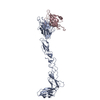
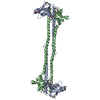
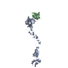
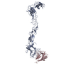
 Map data Map data | This is a twofold-averaged map of the complex formed between dimeric CD28 and the mitogenic antibody 5.11A1. | |||||||||
 Sample Sample |
| |||||||||
| Biological species |   Homo sapiens (human) Homo sapiens (human) | |||||||||
| Method | single particle reconstruction / cryo EM / Resolution: 28.0 Å | |||||||||
 Authors Authors | Evans EJ / Esnouf RM / Manso-Sancho R / Gilbert RJC / James JR / Yu C / Fennely JA / Vowles C / Hanke T / Walse B ...Evans EJ / Esnouf RM / Manso-Sancho R / Gilbert RJC / James JR / Yu C / Fennely JA / Vowles C / Hanke T / Walse B / Hunig T / Sorensen P / Stuart DI / Davis SJ | |||||||||
 Citation Citation | Journal: Nat Immunol / Year: 2005 Title: Crystal structure of a soluble CD28-Fab complex. Authors: Edward J Evans / Robert M Esnouf / Raquel Manso-Sancho / Robert J C Gilbert / John R James / Chao Yu / Janet A Fennelly / Cheryl Vowles / Thomas Hanke / Björn Walse / Thomas Hünig / Poul ...Authors: Edward J Evans / Robert M Esnouf / Raquel Manso-Sancho / Robert J C Gilbert / John R James / Chao Yu / Janet A Fennelly / Cheryl Vowles / Thomas Hanke / Björn Walse / Thomas Hünig / Poul Sørensen / David I Stuart / Simon J Davis /  Abstract: Naive T cell activation requires signaling by the T cell receptor and by nonclonotypic cell surface receptors. The most important costimulatory protein is the monovalent homodimer CD28, which ...Naive T cell activation requires signaling by the T cell receptor and by nonclonotypic cell surface receptors. The most important costimulatory protein is the monovalent homodimer CD28, which interacts with CD80 and CD86 expressed on antigen-presenting cells. Here we present the crystal structure of a soluble form of CD28 in complex with the Fab fragment of a mitogenic antibody. Structural comparisons redefine the evolutionary relationships of CD28-related proteins, antigen receptors and adhesion molecules and account for the distinct ligand-binding and stoichiometric properties of CD28 and the related, inhibitory homodimer CTLA-4. Cryo-electron microscopy-based comparisons of complexes of CD28 with mitogenic and nonmitogenic antibodies place new constraints on models of antibody-induced receptor triggering. This work completes the initial structural characterization of the CD28-CTLA-4-CD80-CD86 signaling system. | |||||||||
| History |
|
- Structure visualization
| Movie |
 Movie viewer Movie viewer |
|---|---|
| Structure viewer | EM map: SurfViewMolmilJmol/JSmol |
| Supplemental images |

- Downloads & links
-EMDB archive
| Map data | emd_1219.map.gz | 204.3 KB | EMDB map data format | |
|---|---|---|---|---|
| Header (meta data) | emd-1219-v30.xmlemd-1219.xml | 11.3 KB 11.3 KB | Display Display | EMDB header |
| Images |  1219.gif 1219.gif | 47.7 KB | ||
| Archive directory |  http://ftp.pdbj.org/pub/emdb/structures/EMD-1219ftp://ftp.pdbj.org/pub/emdb/structures/EMD-1219 http://ftp.pdbj.org/pub/emdb/structures/EMD-1219ftp://ftp.pdbj.org/pub/emdb/structures/EMD-1219 | HTTPS FTP |
-Related structure data


-Links
| EMDB pages | EMDB (EBI/PDBe) / EMDataResource |
|---|
-Map
| File | Download / File: emd_1219.map.gz / Format: CCP4 / Size: 7.8 MB / Type: IMAGE STORED AS FLOATING POINT NUMBER (4 BYTES) | ||||||||||||||||||||||||||||||||||||||||||||||||||||||||||||||||||||
|---|---|---|---|---|---|---|---|---|---|---|---|---|---|---|---|---|---|---|---|---|---|---|---|---|---|---|---|---|---|---|---|---|---|---|---|---|---|---|---|---|---|---|---|---|---|---|---|---|---|---|---|---|---|---|---|---|---|---|---|---|---|---|---|---|---|---|---|---|---|
| Annotation | This is a twofold-averaged map of the complex formed between dimeric CD28 and the mitogenic antibody 5.11A1. | ||||||||||||||||||||||||||||||||||||||||||||||||||||||||||||||||||||
| Projections & slices | Image control
Images are generated by Spider. | ||||||||||||||||||||||||||||||||||||||||||||||||||||||||||||||||||||
| Voxel size | X=Y=Z: 2.60063 Å | ||||||||||||||||||||||||||||||||||||||||||||||||||||||||||||||||||||
| Density |
| ||||||||||||||||||||||||||||||||||||||||||||||||||||||||||||||||||||
| Symmetry | Space group: 1 | ||||||||||||||||||||||||||||||||||||||||||||||||||||||||||||||||||||
| Details | EMDB XML:
CCP4 map header:
| ||||||||||||||||||||||||||||||||||||||||||||||||||||||||||||||||||||
 Z (Sec.)
 Y (Row.)
 X (Col.)


-Supplemental data
- Sample components
-Entire : CD28-Fc fusion complexed with 5.11A1 monoclonal antibody
| Entire | Name: CD28-Fc fusion complexed with 5.11A1 monoclonal antibody |
|---|---|
| Components |
|
-Supramolecule #1000: CD28-Fc fusion complexed with 5.11A1 monoclonal antibody
| Supramolecule | Name: CD28-Fc fusion complexed with 5.11A1 monoclonal antibody type: sample / ID: 1000 Details: The monoclonal antibody 5.11A1 cross links CD28Fc dimers resulting in the formation of filaments. The assymetric unit of the filament is therefore one molecule of CD28Fc and one of Ab 5.11A1. ...Details: The monoclonal antibody 5.11A1 cross links CD28Fc dimers resulting in the formation of filaments. The assymetric unit of the filament is therefore one molecule of CD28Fc and one of Ab 5.11A1. The whole structure presented is the twofold averaged dimer Fab5.11A1-CD28Fc-CD28Fc-Fab5.11A1. Oligomeric state: Filaments of the repeating unit -5.11A1-CD28Fc- Number unique components: 2 |
|---|---|
| Molecular weight | Experimental: 80 KDa / Theoretical: 80 KDa / Method: By sequence and knowledge of oligomeric assembly. |
-Macromolecule #1: CD28Fc fusion
| Macromolecule | Name: CD28Fc fusion / type: protein_or_peptide / ID: 1 / Name.synonym: CD28Fc / Number of copies: 2 / Oligomeric state: Dimer / Recombinant expression: Yes |
|---|---|
| Source (natural) | Organism: Homo sapiens (human) / synonym: Human / Location in cell: Plasma membrane |
| Molecular weight | Experimental: 20 KDa / Theoretical: 20 KDa |
| Recombinant expression | Organism: Lec3.2.8.1 |
-Macromolecule #2: Antibody 5.11A1
| Macromolecule | Name: Antibody 5.11A1 / type: protein_or_peptide / ID: 2 / Name.synonym: 5.11A1 / Number of copies: 2 / Oligomeric state: Dimer / Recombinant expression: No |
|---|---|
| Source (natural) | Organism: Homo sapiens (human) / synonym: Human |
| Molecular weight | Experimental: 60 KDa / Theoretical: 60 KDa |
-Experimental details
-Structure determination
| Method | cryo EM |
|---|---|
 Processing Processing | single particle reconstruction |
| Aggregation state | particle |
-Sample preparation
| Concentration | 0.04 mg/mL |
|---|---|
| Buffer | pH: 7 / Details: HBS |
| Grid | Details: 200 mesh lacey carbon film |
| Vitrification | Cryogen name: ETHANE |
- Electron microscopy
| Microscope | FEI/PHILIPS CM200FEG |
|---|---|
| Electron beam | Acceleration voltage: 200 kV / Electron source: FIELD EMISSION GUN |
| Electron optics | Illumination mode: SPOT SCAN / Imaging mode: BRIGHT FIELDBright-field microscopy / Cs: 2 mm / Nominal defocus max: 7.0 µm / Nominal defocus min: 2.0 µm / Nominal magnification: 50000 |
| Sample stage | Specimen holder: Eucentric / Specimen holder model: GATAN LIQUID NITROGEN |
| Alignment procedure | Legacy - Astigmatism: Corrected at 100,000 X magnification |
| Date | Jan 2, 2004 |
| Image recording | Category: FILM / Film or detector model: KODAK SO-163 FILM / Digitization - Scanner: OTHER / Digitization - Sampling interval: 8.322 µm / Number real images: 10 / Od range: 5 / Bits/pixel: 8 |
-Image processing
| CTF correction | Details: Each micrograph |
|---|---|
| Final reconstruction | Applied symmetry - Point group: C2 (2 fold cyclic) / Algorithm: OTHER / Resolution.type: BY AUTHOR / Resolution: 28.0 Å / Resolution method: FSC 0.5 CUT-OFF / Software - Name: FREALIGN and GAP Details: 3D reconstructions were carried out in FREALIGN without symmetry being imposed and subsequently the twofold symmetry was determined and refined in GAP. Number images used: 529 |
-Atomic model buiding 1
| Initial model | PDB ID: |
|---|---|
| Software | Name: URO |
| Details | Protocol: Rigid body |
| Refinement | Space: REAL / Protocol: RIGID BODY FIT / Target criteria: CC and R-factor |
