Movie
Movie Controller

[English] 日本語
 Yorodumi
- EMDB-11554: Cryo-EM structure of the entire Human topoisomerase II alpha in S... -
+ Open data

- Basic information
| Entry | Database: EMDB / ID: EMD-11554 | ||||||||||||
|---|---|---|---|---|---|---|---|---|---|---|---|---|---|
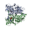
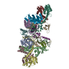
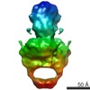
| Title | Cryo-EM structure of the entire Human topoisomerase II alpha in State 2 | ||||||||||||
 Map data Map data | |||||||||||||
 Sample Sample |
| ||||||||||||
 Keywords Keywords |  Human Topoisomerase / Etoposide / DNA / ISOMERASE Human Topoisomerase / Etoposide / DNA / ISOMERASE | ||||||||||||
| Function / homology |  Function and homology information Function and homology informationnegative regulation of DNA duplex unwinding / positive regulation of single stranded viral RNA replication via double stranded DNA intermediate / sister chromatid segregation / apoptotic chromosome condensation / DNA topoisomerase type II (double strand cut, ATP-hydrolyzing) complex / resolution of meiotic recombination intermediates / female meiotic nuclear division / embryonic cleavage / DNA ligation / Transcription of E2F targets under negative control by DREAM complex ...negative regulation of DNA duplex unwinding / positive regulation of single stranded viral RNA replication via double stranded DNA intermediate / sister chromatid segregation / apoptotic chromosome condensation / DNA topoisomerase type II (double strand cut, ATP-hydrolyzing) complex / resolution of meiotic recombination intermediates / female meiotic nuclear division / embryonic cleavage / DNA ligation / Transcription of E2F targets under negative control by DREAM complex / DNA topoisomerase type II (double strand cut, ATP-hydrolyzing) activity / DNA topoisomerase (ATP-hydrolysing) / DNA binding, bending / DNA topological change / SUMOylation of DNA replication proteins / chromosome, centromeric region / ATP-dependent activity, acting on DNA / hematopoietic progenitor cell differentiation / condensed chromosome / protein kinase C binding / ubiquitin binding / male germ cell nucleus / chromosome segregation / regulation of circadian rhythm / rhythmic process / ribonucleoprotein complex / positive regulation of apoptotic process / protein heterodimerization activity / DNA damage response / chromatin binding / nucleolus / magnesium ion binding / protein homodimerization activity / positive regulation of transcription by RNA polymerase II / protein-containing complex / DNA binding / RNA binding / nucleoplasm / ATP binding / nucleus / cytoplasmSimilarity search - Function | ||||||||||||
| Biological species |  Homo sapiens (human) Homo sapiens (human) | ||||||||||||
| Method | single particle reconstruction / cryo EM / Resolution: 7.4 Å | ||||||||||||
 Authors Authors | Vanden Broeck A / Lamour V | ||||||||||||
| Funding support |  France, 3 items France, 3 items
| ||||||||||||
 Citation Citation | Journal: Nat Commun / Year: 2021 Title: Structural basis for allosteric regulation of Human Topoisomerase IIα. Authors: Arnaud Vanden Broeck / Christophe Lotz / Robert Drillien / Léa Haas / Claire Bedez / Valérie Lamour / Abstract: The human type IIA topoisomerases (Top2) are essential enzymes that regulate DNA topology and chromosome organization. The Topo IIα isoform is a prime target for antineoplastic compounds used in ...The human type IIA topoisomerases (Top2) are essential enzymes that regulate DNA topology and chromosome organization. The Topo IIα isoform is a prime target for antineoplastic compounds used in cancer therapy that form ternary cleavage complexes with the DNA. Despite extensive studies, structural information on this large dimeric assembly is limited to the catalytic domains, hindering the exploration of allosteric mechanism governing the enzyme activities and the contribution of its non-conserved C-terminal domain (CTD). Herein we present cryo-EM structures of the entire human Topo IIα nucleoprotein complex in different conformations solved at subnanometer resolutions (3.6-7.4 Å). Our data unveils the molecular determinants that fine tune the allosteric connections between the ATPase domain and the DNA binding/cleavage domain. Strikingly, the reconstruction of the DNA-binding/cleavage domain uncovers a linker leading to the CTD, which plays a critical role in modulating the enzyme's activities and opens perspective for the analysis of post-translational modifications. | ||||||||||||
| History |
|
- Structure visualization
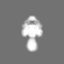
| Movie |
Movie viewer |
|---|---|
| Structure viewer | EM map: SurfViewMolmilJmol/JSmol |
| Supplemental images |

- Downloads & links
-EMDB archive
| Map data | emd_11554.map.gz | 36.4 MB | EMDB map data format | |
|---|---|---|---|---|
| Header (meta data) | emd-11554-v30.xmlemd-11554.xml | 21.5 KB 21.5 KB | Display Display | EMDB header |
| FSC (resolution estimation) | emd_11554_fsc.xml | 10 KB | Display | FSC data file |
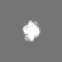
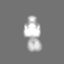
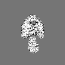
| Images | 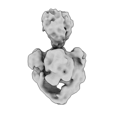 emd_11554.png emd_11554.png | 36.5 KB | ||
| Masks | emd_11554_msk_1.map | 38.4 MB | Mask map | |
| Filedesc metadata | emd-11554.cif.gz | 7.3 KB | ||
| Others | emd_11554_half_map_1.map.gzemd_11554_half_map_2.map.gz | 3.2 MB 3.2 MB | ||
| Archive directory |  http://ftp.pdbj.org/pub/emdb/structures/EMD-11554ftp://ftp.pdbj.org/pub/emdb/structures/EMD-11554 http://ftp.pdbj.org/pub/emdb/structures/EMD-11554ftp://ftp.pdbj.org/pub/emdb/structures/EMD-11554 | HTTPS FTP |
-Related structure data
| Related structure data |  6zy8MC  6zy5C 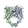 6zy6C  6zy7C M: atomic model generated by this map C: citing same article ( |
|---|---|
| Similar structure data |


-Links
| EMDB pages | EMDB (EBI/PDBe) / EMDataResource |
|---|---|
| Related items in Molecule of the Month |


-Map
| File | Download / File: emd_11554.map.gz / Format: CCP4 / Size: 38.4 MB / Type: IMAGE STORED AS FLOATING POINT NUMBER (4 BYTES) | ||||||||||||||||||||||||||||||||||||||||||||||||||||||||||||
|---|---|---|---|---|---|---|---|---|---|---|---|---|---|---|---|---|---|---|---|---|---|---|---|---|---|---|---|---|---|---|---|---|---|---|---|---|---|---|---|---|---|---|---|---|---|---|---|---|---|---|---|---|---|---|---|---|---|---|---|---|---|
| Voxel size | X=Y=Z: 1.507 Å | ||||||||||||||||||||||||||||||||||||||||||||||||||||||||||||
| Density |
| ||||||||||||||||||||||||||||||||||||||||||||||||||||||||||||
| Symmetry | Space group: 1 | ||||||||||||||||||||||||||||||||||||||||||||||||||||||||||||
| Details | EMDB XML:
CCP4 map header:
| ||||||||||||||||||||||||||||||||||||||||||||||||||||||||||||
-Supplemental data
-Mask #1
| File | emd_11554_msk_1.map | ||||||||||||
|---|---|---|---|---|---|---|---|---|---|---|---|---|---|
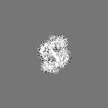
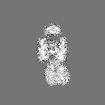
| Projections & Slices |
| ||||||||||||
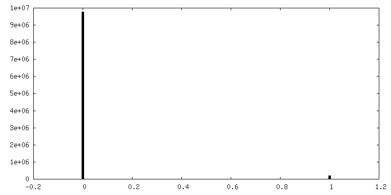
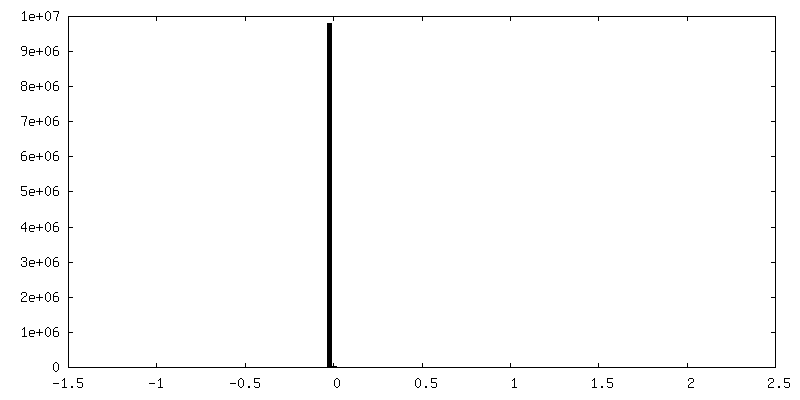
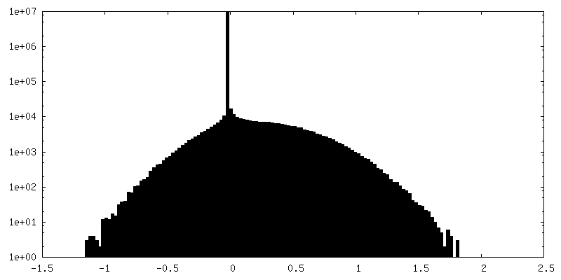
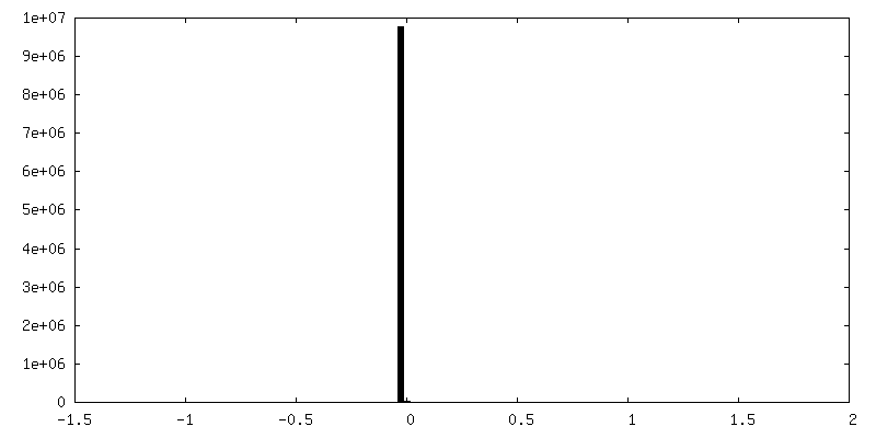
| Density Histograms |
 Z
Z Y
Y X
X


-Half map: #2
| File | emd_11554_half_map_1.map | ||||||||||||
|---|---|---|---|---|---|---|---|---|---|---|---|---|---|
| Projections & Slices |
| ||||||||||||
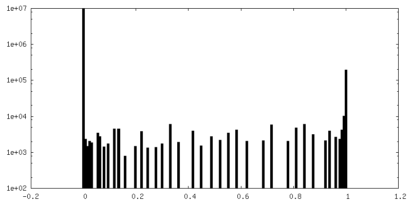
| Density Histograms |

-Half map: #1
| File | emd_11554_half_map_2.map | ||||||||||||
|---|---|---|---|---|---|---|---|---|---|---|---|---|---|
| Projections & Slices |
| ||||||||||||
| Density Histograms |


- Sample components
-Entire : Etoposide-bound Entire Human Top2a in state 2
| Entire | Name: Etoposide-bound Entire Human Top2a in state 2 |
|---|---|
| Components |
|
-Supramolecule #1: Etoposide-bound Entire Human Top2a in state 2
| Supramolecule | Name: Etoposide-bound Entire Human Top2a in state 2 / type: complex / ID: 1 / Parent: 0 / Macromolecule list: #1-#3 |
|---|---|
| Molecular weight | Theoretical: 295 KDa |
-Supramolecule #2: DNA topoisomerase 2-alpha
| Supramolecule | Name: DNA topoisomerase 2-alpha / type: complex / ID: 2 / Parent: 1 / Macromolecule list: #1 |
|---|---|
| Source (natural) | Organism: Homo sapiens (human) |
-Supramolecule #3: DNA
| Supramolecule | Name: DNA / type: complex / ID: 3 / Parent: 1 / Macromolecule list: #2-#3 |
|---|---|
| Source (natural) | Organism: Homo sapiens (human) |
-Macromolecule #1: DNA topoisomerase 2-alpha
| Macromolecule | Name: DNA topoisomerase 2-alpha / type: protein_or_peptide / ID: 1 / Number of copies: 2 / Enantiomer: LEVO / EC number: DNA topoisomerase (ATP-hydrolysing) |
|---|---|
| Source (natural) | Organism: Homo sapiens (human) |
| Molecular weight | Theoretical: 174.654406 KDa |
| Recombinant expression | Organism:  Vaccinia virus Ankara Vaccinia virus Ankara |
| Sequence | String: MEVSPLQPVN ENMQVNKIKK NEDAKKRLSV ERIYQKKTQL EHILLRPDTY IGSVELVTQQ MWVYDEDVGI NYREVTFVPG LYKIFDEIL VNAADNKQRD PKMSCIRVTI DPENNLISIW NNGKGIPVVE HKVEKMYVPA LIFGQLLTSS NYDDDEKKVT G GRNGYGAK ...String: MEVSPLQPVN ENMQVNKIKK NEDAKKRLSV ERIYQKKTQL EHILLRPDTY IGSVELVTQQ MWVYDEDVGI NYREVTFVPG LYKIFDEIL VNAADNKQRD PKMSCIRVTI DPENNLISIW NNGKGIPVVE HKVEKMYVPA LIFGQLLTSS NYDDDEKKVT G GRNGYGAK LCNIFSTKFT VETASREYKK MFKQTWMDNM GRAGEMELKP FNGEDYTCIT FQPDLSKFKM QSLDKDIVAL MV RRAYDIA GSTKDVKVFL NGNKLPVKGF RSYVDMYLKD KLDETGNSLK VIHEQVNHRW EVCLTMSEKG FQQISFVNSI ATS KGGRHV DYVADQIVTK LVDVVKKKNK GGVAVKAHQV KNHMWIFVNA LIENPTFDSQ TKENMTLQPK SFGSTCQLSE KFIK AAIGC GIVESILNWV KFKAQVQLNK KCSAVKHNRI KGIPKLDDAN DAGGRNSTEC TLILTEGDSA KTLAVSGLGV VGRDK YGVF PLRGKILNVR EASHKQIMEN AEINNIIKIV GLQYKKNYED EDSLKTLRYG KIMIMTDQDQ DGSHIKGLLI NFIHHN WPS LLRHRFLEEF ITPIVKVSKN KQEMAFYSLP EFEEWKSSTP NHKKWKVKYY KGLGTSTSKE AKEYFADMKR HRIQFKY SG PEDDAAISLA FSKKQIDDRK EWLTNFMEDR RQRKLLGLPE DYLYGQTTTY LTYNDFINKE LILFSNSDNE RSIPSMVD G LKPGQRKVLF TCFKRNDKRE VKVAQLAGSV AEMSSYHHGE MSLMMTIINL AQNFVGSNNL NLLQPIGQFG TRLHGGKDS ASPRYIFTML SSLARLLFPP KDDHTLKFLY DDNQRVEPEW YIPIIPMVLI NGAEGIGTGW SCKIPNFDVR EIVNNIRRLM DGEEPLPML PSYKNFKGTI EELAPNQYVI SGEVAILNST TIEISELPVR TWTQTYKEQV LEPMLNGTEK TPPLITDYRE Y HTDTTVKF VVKMTEEKLA EAERVGLHKV FKLQTSLTCN SMVLFDHVGC LKKYDTVLDI LRDFFELRLK YYGLRKEWLL GM LGAESAK LNNQARFILE KIDGKIIIEN KPKKELIKVL IQRGYDSDPV KAWKEAQQKV PDEEENEESD NEKETEKSDS VTD SGPTFN YLLDMPLWYL TKEKKDELCR LRNEKEQELD TLKRKSPSDL WKEDLATFIE ELEAVEAKEK QDEQVGLPGK GGKA KGKKT QMAEVLPSPR GQRVIPRITI EMKAEAEKKN KKKIKNENTE GSPQEDGVEL EGLKQRLEKK QKREPGTKTK KQTTL AFKP IKKGKKRNPW SDSESDRSSD ESNFDVPPRE TEPRRAATKT KFTMDLDSDE DFSDFDEKTD DEDFVPSDAS PPKTKT SPK LSNKELKPQK SVVSDLEADD VKGSVPLSSS PPATHFPDET EITNPVPKKN VTVKKTAAKS QSSTSTTGAK KRAAPKG TK RDPALNSGVS QKPDPAKTKN RRKRKPSTSD DSDSNFEKIV SKAVTSKKSK GESDDFHMDF DSAVAPRAKS VRAKKPIK Y LEESDEDDLF UniProtKB: DNA topoisomerase 2-alpha |
-Macromolecule #2: DNA (5'-D(*CP*GP*CP*GP*CP*AP*TP*CP*GP*TP*CP*AP*TP*CP*CP*TP*C)-3')
| Macromolecule | Name: DNA (5'-D(*CP*GP*CP*GP*CP*AP*TP*CP*GP*TP*CP*AP*TP*CP*CP*TP*C)-3') type: dna / ID: 2 / Number of copies: 2 / Classification: DNA |
|---|---|
| Source (natural) | Organism: Homo sapiens (human) |
| Molecular weight | Theoretical: 5.099298 KDa |
| Sequence | String: (DC)(DG)(DC)(DG)(DC)(DA)(DT)(DC)(DG)(DT) (DC)(DA)(DT)(DC)(DC)(DT)(DC) |
-Macromolecule #3: DNA (5'-D(*GP*AP*GP*GP*AP*TP*GP*AP*CP*GP*AP*TP*G)-3')
| Macromolecule | Name: DNA (5'-D(*GP*AP*GP*GP*AP*TP*GP*AP*CP*GP*AP*TP*G)-3') / type: dna / ID: 3 / Number of copies: 2 / Classification: DNA |
|---|---|
| Source (natural) | Organism: Homo sapiens (human) |
| Molecular weight | Theoretical: 4.080671 KDa |
| Sequence | String: (DG)(DA)(DG)(DG)(DA)(DT)(DG)(DA)(DC)(DG) (DA)(DT)(DG) |
-Macromolecule #4: PHOSPHOAMINOPHOSPHONIC ACID-ADENYLATE ESTER
| Macromolecule | Name: PHOSPHOAMINOPHOSPHONIC ACID-ADENYLATE ESTER / type: ligand / ID: 4 / Number of copies: 2 / Formula: ANP |
|---|---|
| Molecular weight | Theoretical: 506.196 Da |
| Chemical component information |  ChemComp-ANP: |
-Macromolecule #5: (5S,5aR,8aR,9R)-9-(4-hydroxy-3,5-dimethoxyphenyl)-8-oxo-5,5a,6,8,...
| Macromolecule | Name: (5S,5aR,8aR,9R)-9-(4-hydroxy-3,5-dimethoxyphenyl)-8-oxo-5,5a,6,8,8a,9-hexahydrofuro[3',4':6,7]naphtho[2,3-d][1,3]dioxol -5-yl 4,6-O-[(1R)-ethylidene]-beta-D-glucopyranoside type: ligand / ID: 5 / Number of copies: 2 / Formula: EVP |
|---|---|
| Molecular weight | Theoretical: 588.557 Da |
-Experimental details
-Structure determination
| Method | cryo EM |
|---|---|
 Processing Processing | single particle reconstruction |
| Aggregation state | particle |
-Sample preparation
| Buffer | pH: 8 |
|---|---|
| Vitrification | Cryogen name: ETHANE / Chamber humidity: 95 % / Chamber temperature: 283 K / Instrument: FEI VITROBOT MARK IV |
- Electron microscopy
| Microscope | FEI TITAN KRIOS |
|---|---|
| Electron beam | Acceleration voltage: 300 kV / Electron source: FIELD EMISSION GUN |
| Electron optics | Illumination mode: FLOOD BEAM / Imaging mode: BRIGHT FIELDBright-field microscopy / Cs: 0.01 mm / Nominal defocus max: 3.0 µm / Nominal defocus min: 1.0 µm / Nominal magnification: 105000 |
| Specialist optics | Energy filter - Name: GIF Quantum LS / Energy filter - Slit width: 20 eV |
| Sample stage | Specimen holder model: FEI TITAN KRIOS AUTOGRID HOLDER / Cooling holder cryogen: NITROGEN |
| Image recording | Film or detector model: GATAN K2 SUMMIT (4k x 4k) / Detector mode: SUPER-RESOLUTION / Average electron dose: 50.0 e/Å2 |
| Experimental equipment |  Model: Titan Krios / Image courtesy: FEI Company |
-Image processing
| Particle selection | Number selected: 1908092 |
|---|---|
| Startup model | #0 - Type of model: INSILICO MODEL #0 - In silico model: An ab initio model was reconstructed using cryoSPARC #1 - Type of model: INSILICO MODEL #1 - In silico model: An ab initio model was reconstructed using cryoSPARC |
| Initial angle assignment | Type: MAXIMUM LIKELIHOOD / Software - Name: cryoSPARC (ver. v3) |
| Final angle assignment | Type: MAXIMUM LIKELIHOOD / Software - Name: cryoSPARC (ver. v3) |
| Final reconstruction | Applied symmetry - Point group: C1 (asymmetric) / Resolution.type: BY AUTHOR / Resolution: 7.4 Å / Resolution method: FSC 0.143 CUT-OFF / Software - Name: cryoSPARC (ver. v3) / Number images used: 13420 |
| FSC plot (resolution estimation) | 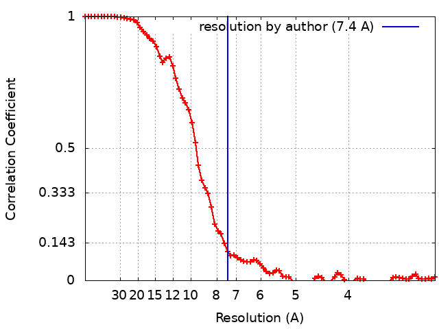 |