 Movie
Movie Controller

 Yorodumi
+ Open data

- Basic information
| Entry | Database: EMDB / ID: EMD-1143 | |||||||||
|---|---|---|---|---|---|---|---|---|---|---|
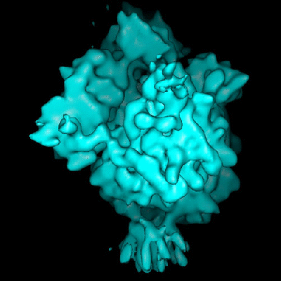
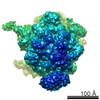
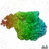
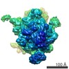
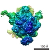
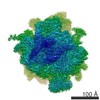
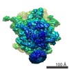
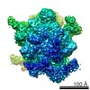
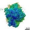
| Title | Structure of the E. coli protein-conducting channel bound to a translating ribosome. | |||||||||
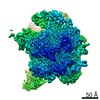
 Map data Map data | EM map of the E.coli proten-conducting channel bound to a translating ribosome | |||||||||
 Sample Sample |
| |||||||||
| Function / homology |  Function and homology information Function and homology informationprotein insertion into membrane from inner side / cell envelope Sec protein transport complex / protein transport by the Sec complex /  intracellular protein transmembrane transport / protein-transporting ATPase activity / SRP-dependent cotranslational protein targeting to membrane, translocation / signal sequence binding / stringent response / mRNA base-pairing translational repressor activity / ornithine decarboxylase inhibitor activity ...protein insertion into membrane from inner side / cell envelope Sec protein transport complex / protein transport by the Sec complex / intracellular protein transmembrane transport / protein-transporting ATPase activity / SRP-dependent cotranslational protein targeting to membrane, translocation / signal sequence binding / stringent response / mRNA base-pairing translational repressor activity / ornithine decarboxylase inhibitor activity / misfolded RNA binding / transcription antitermination factor activity, RNA binding / Group I intron splicing / RNA folding / protein transmembrane transporter activity / protein secretion / protein targeting / transcriptional attenuation / endoribonuclease inhibitor activity / RNA-binding transcription regulator activity / positive regulation of ribosome biogenesis / negative regulation of cytoplasmic translation / translational termination / DnaA-L2 complex / four-way junction DNA binding / translation repressor activity / negative regulation of translational initiation / negative regulation of DNA-templated DNA replication initiation / regulation of mRNA stability / ribosome assembly / mRNA regulatory element binding translation repressor activity / response to reactive oxygen species / assembly of large subunit precursor of preribosome / transcription elongation factor complex / positive regulation of RNA splicing / DNA endonuclease activity / : / cytosolic ribosome assembly / regulation of DNA-templated transcription elongation / transcription antitermination / regulation of cell growth / maintenance of translational fidelity / DNA-templated transcription termination / intracellular protein transport / response to radiation / mRNA 5'-UTR binding / ribosomal small subunit biogenesis / small ribosomal subunit rRNA binding / ribosomal small subunit assembly / ribosomal large subunit assembly / cytosolic small ribosomal subunit / large ribosomal subunit rRNA binding / ribosome binding / large ribosomal subunit / ribosome biogenesis / regulation of translation / small ribosomal subunit / 5S rRNA binding / cytosolic large ribosomal subunit / cytoplasmic translation / transferase activity / tRNA binding / negative regulation of translation / membrane => GO:0016020 / rRNA binding / molecular adaptor activity / ribosome / structural constituent of ribosome / translation / response to antibiotic / mRNA binding / negative regulation of DNA-templated transcription / protein homodimerization activity / DNA binding / RNA binding / zinc ion binding / membrane / plasma membrane / cytosol / cytoplasm intracellular protein transmembrane transport / protein-transporting ATPase activity / SRP-dependent cotranslational protein targeting to membrane, translocation / signal sequence binding / stringent response / mRNA base-pairing translational repressor activity / ornithine decarboxylase inhibitor activity ...protein insertion into membrane from inner side / cell envelope Sec protein transport complex / protein transport by the Sec complex / intracellular protein transmembrane transport / protein-transporting ATPase activity / SRP-dependent cotranslational protein targeting to membrane, translocation / signal sequence binding / stringent response / mRNA base-pairing translational repressor activity / ornithine decarboxylase inhibitor activity / misfolded RNA binding / transcription antitermination factor activity, RNA binding / Group I intron splicing / RNA folding / protein transmembrane transporter activity / protein secretion / protein targeting / transcriptional attenuation / endoribonuclease inhibitor activity / RNA-binding transcription regulator activity / positive regulation of ribosome biogenesis / negative regulation of cytoplasmic translation / translational termination / DnaA-L2 complex / four-way junction DNA binding / translation repressor activity / negative regulation of translational initiation / negative regulation of DNA-templated DNA replication initiation / regulation of mRNA stability / ribosome assembly / mRNA regulatory element binding translation repressor activity / response to reactive oxygen species / assembly of large subunit precursor of preribosome / transcription elongation factor complex / positive regulation of RNA splicing / DNA endonuclease activity / : / cytosolic ribosome assembly / regulation of DNA-templated transcription elongation / transcription antitermination / regulation of cell growth / maintenance of translational fidelity / DNA-templated transcription termination / intracellular protein transport / response to radiation / mRNA 5'-UTR binding / ribosomal small subunit biogenesis / small ribosomal subunit rRNA binding / ribosomal small subunit assembly / ribosomal large subunit assembly / cytosolic small ribosomal subunit / large ribosomal subunit rRNA binding / ribosome binding / large ribosomal subunit / ribosome biogenesis / regulation of translation / small ribosomal subunit / 5S rRNA binding / cytosolic large ribosomal subunit / cytoplasmic translation / transferase activity / tRNA binding / negative regulation of translation / membrane => GO:0016020 / rRNA binding / molecular adaptor activity / ribosome / structural constituent of ribosome / translation / response to antibiotic / mRNA binding / negative regulation of DNA-templated transcription / protein homodimerization activity / DNA binding / RNA binding / zinc ion binding / membrane / plasma membrane / cytosol / cytoplasmSimilarity search - Function | |||||||||
| Biological species |  Escherichia coli (E. coli) / unidentified (others) Escherichia coli (E. coli) / unidentified (others) | |||||||||
| Method | single particle reconstruction / cryo EM / Resolution: 14.9 Å | |||||||||
 Authors Authors | Mitra K / Schaffitzel C / Shaikh T / Tama F / Jenni S / Brooks III CL / Ban N / Frank J | |||||||||
 Citation Citation | Journal: Nature / Year: 2005 Title: Structure of the E. coli protein-conducting channel bound to a translating ribosome. Authors: Kakoli Mitra / Christiane Schaffitzel / Tanvir Shaikh / Florence Tama / Simon Jenni / Charles L Brooks / Nenad Ban / Joachim Frank /  Abstract: Secreted and membrane proteins are translocated across or into cell membranes through a protein-conducting channel (PCC). Here we present a cryo-electron microscopy reconstruction of the Escherichia ...Secreted and membrane proteins are translocated across or into cell membranes through a protein-conducting channel (PCC). Here we present a cryo-electron microscopy reconstruction of the Escherichia coli PCC, SecYEG, complexed with the ribosome and a nascent chain containing a signal anchor. This reconstruction shows a messenger RNA, three transfer RNAs, the nascent chain, and detailed features of both a translocating PCC and a second, non-translocating PCC bound to mRNA hairpins. The translocating PCC forms connections with ribosomal RNA hairpins on two sides and ribosomal proteins at the back, leaving a frontal opening. Normal mode-based flexible fitting of the archaeal SecYEbeta structure into the PCC electron microscopy densities favours a front-to-front arrangement of two SecYEG complexes in the PCC, and supports channel formation by the opening of two linked SecY halves during polypeptide translocation. On the basis of our observation in the translocating PCC of two segregated pores with different degrees of access to bulk lipid, we propose a model for co-translational protein translocation. | |||||||||
| History |
|
- Structure visualization
| Movie |
Movie viewer |
|---|---|
| Structure viewer | EM map: SurfViewMolmilJmol/JSmol |
| Supplemental images |
- Downloads & links
-EMDB archive
| Map data | emd_1143.map.gz | 6.2 MB | EMDB map data format | |
|---|---|---|---|---|
| Header (meta data) | emd-1143-v30.xmlemd-1143.xml | 14.3 KB 14.3 KB | Display Display | EMDB header |
| Images |  1143.gif 1143.gif | 67.6 KB | ||
| Archive directory |  http://ftp.pdbj.org/pub/emdb/structures/EMD-1143ftp://ftp.pdbj.org/pub/emdb/structures/EMD-1143 http://ftp.pdbj.org/pub/emdb/structures/EMD-1143ftp://ftp.pdbj.org/pub/emdb/structures/EMD-1143 | HTTPS FTP |
-Related structure data
| Related structure data |  2akhMC  2akiMC  4v4wM M: atomic model generated by this map C: citing same article ( |
|---|---|
| Similar structure data |

-Links
| EMDB pages | EMDB (EBI/PDBe) / EMDataResource |
|---|---|
| Related items in Molecule of the Month |


-Map
| File | Download / File: emd_1143.map.gz / Format: CCP4 / Size: 6.6 MB / Type: IMAGE STORED AS FLOATING POINT NUMBER (4 BYTES) | ||||||||||||||||||||||||||||||||||||||||||||||||||||||||||||||||||||
|---|---|---|---|---|---|---|---|---|---|---|---|---|---|---|---|---|---|---|---|---|---|---|---|---|---|---|---|---|---|---|---|---|---|---|---|---|---|---|---|---|---|---|---|---|---|---|---|---|---|---|---|---|---|---|---|---|---|---|---|---|---|---|---|---|---|---|---|---|---|
| Annotation | EM map of the E.coli proten-conducting channel bound to a translating ribosome | ||||||||||||||||||||||||||||||||||||||||||||||||||||||||||||||||||||
| Voxel size |
| ||||||||||||||||||||||||||||||||||||||||||||||||||||||||||||||||||||
| Density |
| ||||||||||||||||||||||||||||||||||||||||||||||||||||||||||||||||||||
| Symmetry | Space group: 1 | ||||||||||||||||||||||||||||||||||||||||||||||||||||||||||||||||||||
| Details | EMDB XML:
CCP4 map header:
| ||||||||||||||||||||||||||||||||||||||||||||||||||||||||||||||||||||
-Supplemental data
- Sample components
-Entire : co-translational E. coli 70S ribosome-nascent chain complexed wit...
| Entire | Name: co-translational E. coli 70S ribosome-nascent chain complexed with SecYEG |
|---|---|
| Components |
|
-Supramolecule #1000: co-translational E. coli 70S ribosome-nascent chain complexed wit...
| Supramolecule | Name: co-translational E. coli 70S ribosome-nascent chain complexed with SecYEG type: sample / ID: 1000 / Number unique components: 8 |
|---|
-Supramolecule #1: 30S
| Supramolecule | Name: 30S / type: complex / ID: 1 / Name.synonym: small subunit / Recombinant expression: No / Ribosome-details: ribosome-prokaryote: SSU 30S |
|---|---|
| Source (natural) | Organism: Escherichia coli (E. coli) |
-Supramolecule #2: 50S
| Supramolecule | Name: 50S / type: complex / ID: 2 / Name.synonym: large subunit / Recombinant expression: No / Ribosome-details: ribosome-prokaryote: LSU 50S |
|---|---|
| Source (natural) | Organism: Escherichia coli (E. coli) |
-Macromolecule #1: A-site tRNA
| Macromolecule | Name: A-site tRNA / type: rna / ID: 1 / Classification: OTHER / Structure: DOUBLE HELIX / Synthetic?: No |
|---|---|
| Source (natural) | Organism: Escherichia coli (E. coli) |
-Macromolecule #2: P-site tRNA
| Macromolecule | Name: P-site tRNA / type: rna / ID: 2 / Classification: OTHER / Structure: DOUBLE HELIX / Synthetic?: No |
|---|---|
| Source (natural) | Organism: Escherichia coli (E. coli) |
-Macromolecule #3: E-site tRNA
| Macromolecule | Name: E-site tRNA / type: rna / ID: 3 / Classification: OTHER / Structure: DOUBLE HELIX / Synthetic?: No |
|---|---|
| Source (natural) | Organism: Escherichia coli (E. coli) |
-Macromolecule #4: mRNA
| Macromolecule | Name: mRNA / type: rna / ID: 4 / Classification: OTHER / Structure: SINGLE STRANDED / Synthetic?: Yes |
|---|---|
| Source (natural) | Organism: Escherichia coli (E. coli) |
-Macromolecule #5: protein-conducting channel
| Macromolecule | Name: protein-conducting channel / type: protein_or_peptide / ID: 5 / Name.synonym: translocon, SecYEG / Number of copies: 2 / Oligomeric state: dimer of heterotrimer / Recombinant expression: Yes |
|---|---|
| Source (natural) | Organism: Escherichia coli (E. coli) / synonym: translocon / Location in cell: inner membrane |
| Recombinant expression | Organism: Escherichia coli (E. coli) |
-Macromolecule #6: nascent chain
| Macromolecule | Name: nascent chain / type: protein_or_peptide / ID: 6 / Details: Strep-II-FtsQ-SecM construct / Recombinant expression: Yes |
|---|---|
| Source (natural) | Organism: unidentified (others) / synonym: translocon |
-Experimental details
-Structure determination
| Method | cryo EM |
|---|---|
 Processing Processing | single particle reconstruction |
| Aggregation state | particle |
-Sample preparation
| Vitrification | Cryogen name: ETHANE / Chamber humidity: 90 % / Chamber temperature: 93 K / Instrument: HOMEMADE PLUNGER Details: Vitrification instrument: two sided blotting plunger Method: Blot for 2 seconds before plunging |
|---|
- Electron microscopy
| Microscope | FEI TECNAI F30 |
|---|---|
| Electron beam | Acceleration voltage: 300 kV / Electron source: FIELD EMISSION GUN |
| Electron optics | Calibrated magnification: 39000 / Illumination mode: FLOOD BEAM / Imaging mode: BRIGHT FIELDBright-field microscopy / Cs: 2.26 mm / Nominal defocus max: 4.3 µm / Nominal defocus min: 1.5 µm / Nominal magnification: 39000 |
| Sample stage | Specimen holder: Cryo stage / Specimen holder model: OTHER |
| Temperature | Average: 93 K |
| Date | Mar 9, 2004 |
| Image recording | Category: FILM / Film or detector model: KODAK SO-163 FILM / Digitization - Scanner: ZEISS SCAI / Digitization - Sampling interval: 14 µm / Number real images: 385 / Average electron dose: 11 e/Å2 / Od range: 1.2 / Bits/pixel: 12 |
| Experimental equipment |  Model: Tecnai F30 / Image courtesy: FEI Company |
-Image processing
| CTF correction | Details: defocus groups |
|---|---|
| Final angle assignment | Details: SPIDER: theta 15 degrees, phi 15 degrees |
| Final reconstruction | Applied symmetry - Point group: C1 (asymmetric) / Algorithm: OTHER / Resolution.type: BY AUTHOR / Resolution: 14.9 Å / Resolution method: FSC 0.5 CUT-OFF / Software - Name: SPIDER package Details: The falloff of Fourier amplitudes toward higher spatial frequencies was corrected using the x-ray solution scattering intensity distribution of 70S ribosomes from E. coli during each round of refinement Number images used: 53325 |
-Atomic model buiding 1
| Details | Protocol: normal mode-based flexible fitting. Fitting of SecYEG atomic model into isolated EM density of protein-conducting channels |
|---|---|
| Refinement | Space: REAL / Protocol: FLEXIBLE FIT / Target criteria: correlation coefficient |
| Output model | PDB-2akh: PDB-2aki: PDB-4v4w: |
-Atomic model buiding 2
| Details | Protocol: normal mode-based flexible fitting. Fitting of SecYEG atomic model into isolated EM density of protein-conducting channels |
|---|---|
| Refinement | Space: REAL / Protocol: FLEXIBLE FIT / Target criteria: correlation coefficient |
| Output model | PDB-2akh: PDB-2aki: PDB-4v4w: |
-Atomic model buiding 3
| Software | Name: RSR2000 |
|---|---|
| Details | Protocol: real space refinement |
| Refinement | Space: REAL / Protocol: RIGID BODY FIT / Target criteria: R-factor |
| Output model | PDB-2akh: PDB-2aki: PDB-4v4w: |
-Atomic model buiding 4
| Software | Name: RSR2000 |
|---|---|
| Details | Protocol: real space refinement |
| Refinement | Space: REAL / Protocol: RIGID BODY FIT / Target criteria: R-factor |
| Output model | PDB-2akh: PDB-2aki: PDB-4v4w: |
