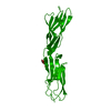
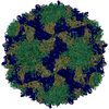
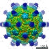
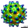
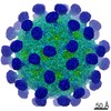
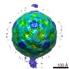
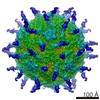
Journal: Structure / Year: 2005 Title: The crystal structure of coxsackievirus A21 and its interaction with ICAM-1. Authors: Chuan Xiao / Carol M Bator-Kelly / Elizabeth Rieder / Paul R Chipman / Alister Craig / Richard J Kuhn / Eckard Wimmer / Michael G Rossmann / Abstract: CVA21 and polioviruses both belong to the Enterovirus genus in the family of Picornaviridae, whereas rhinoviruses form a distinct picornavirus genus. Nevertheless, CVA21 and the major group of human ...CVA21 and polioviruses both belong to the Enterovirus genus in the family of Picornaviridae, whereas rhinoviruses form a distinct picornavirus genus. Nevertheless, CVA21 and the major group of human rhinoviruses recognize intercellular adhesion molecule-1 (ICAM-1) as their cellular receptor, whereas polioviruses use poliovirus receptor. The crystal structure of CVA21 has been determined to 3.2 A resolution. Its structure has greater similarity to poliovirus structures than to other known picornavirus structures. Cryo-electron microscopy (cryo-EM) was used to determine an 8.0 A resolution structure of CVA21 complexed with an ICAM-1 variant, ICAM-1(Kilifi). The cryo-EM map was fitted with the crystal structures of ICAM-1 and CVA21. Significant differences in the structure of CVA21 with respect to the poliovirus structures account for the inability of ICAM-1 to bind polioviruses. The interface between CVA21 and ICAM-1 has shape and electrostatic complementarity with many residues being conserved among those CVAs that bind ICAM-1.
History
Deposition
Mar 29, 2005
-
Header (metadata) release
Mar 29, 2005
-
Map release
Oct 4, 2005
-
Update
May 26, 2011
-
Current status
May 26, 2011
Processing site: PDBe / Status: Released
-
Structure visualization
Movie
Surface view with section colored by density value
Category: FILM / Film or detector model: KODAK SO-163 FILM / Digitization - Scanner: ZEISS SCAI / Digitization - Sampling interval: 2.98 µm / Number real images: 33 / Average electron dose: 30 e/Å2 / Details: 7um step scanned and averaged to 14um / Bits/pixel: 8
Tilt angle min
0
Tilt angle max
0
-
Image processing
CTF correction
Details: Each particle
Final reconstruction
Applied symmetry - Point group: I (icosahedral) / Algorithm: OTHER / Resolution.type: BY AUTHOR / Resolution: 8.0 Å / Resolution method: FSC 0.5 CUT-OFF / Software - Name: parallelized EMPFT, EM3DR / Number images used: 4704
In the structure databanks used in Yorodumi, some data are registered as the other names, "COVID-19 virus" and "2019-nCoV". Here are the details of the virus and the list of structure data.
Jan 31, 2019. EMDB accession codes are about to change! (news from PDBe EMDB page)
EMDB accession codes are about to change! (news from PDBe EMDB page)
The allocation of 4 digits for EMDB accession codes will soon come to an end. Whilst these codes will remain in use, new EMDB accession codes will include an additional digit and will expand incrementally as the available range of codes is exhausted. The current 4-digit format prefixed with “EMD-” (i.e. EMD-XXXX) will advance to a 5-digit format (i.e. EMD-XXXXX), and so on. It is currently estimated that the 4-digit codes will be depleted around Spring 2019, at which point the 5-digit format will come into force.
The EM Navigator/Yorodumi systems omit the EMD- prefix.
Related info.:Q: What is EMD? / ID/Accession-code notation in Yorodumi/EM Navigator
Yorodumi is a browser for structure data from EMDB, PDB, SASBDB, etc.
This page is also the successor to EM Navigator detail page, and also detail information page/front-end page for Omokage search.
The word "yorodu" (or yorozu) is an old Japanese word meaning "ten thousand". "mi" (miru) is to see.
Related info.:EMDB / PDB / SASBDB / Comparison of 3 databanks / Yorodumi Search / Aug 31, 2016. New EM Navigator & Yorodumi / Yorodumi Papers / Jmol/JSmol / Function and homology information / Changes in new EM Navigator and Yorodumi
 Movie
Movie Controller

 Yorodumi
 Open data
 Basic information
 Map data
 Sample
 Function and homology information
 immunological synapse / Integrin cell surface interactions / negative regulation of endothelial cell apoptotic process / negative regulation of extrinsic apoptotic signaling pathway via death domain receptors / cellular response to leukemia inhibitory factor /
 Authors
 Citation
 Structure visualization
 Downloads & links
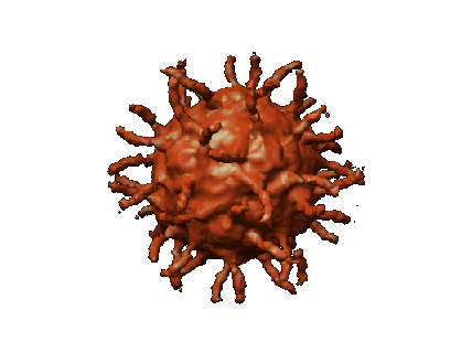 1114.gif
 http://ftp.pdbj.org/pub/emdb/structures/EMD-1114
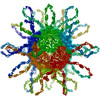


 Sample components
 Processing
 Electron microscopy