 Movie
Movie Controller


+ Open data

- Basic information
| Entry | Database: EMDB / ID: EMD-11023 | |||||||||
|---|---|---|---|---|---|---|---|---|---|---|
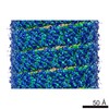
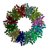
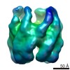
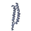
| Title | CryoEM structure of the Chikungunya virus nsP1 complex | |||||||||
 Map data Map data | ||||||||||
 Sample Sample |
| |||||||||
 Keywords Keywords |  Capping enzyme / monotopic membrane complex / membrane bending / membrane pore / replication complex / scaffold / Spherule formation / viral factory. / VIRAL PROTEIN Capping enzyme / monotopic membrane complex / membrane bending / membrane pore / replication complex / scaffold / Spherule formation / viral factory. / VIRAL PROTEIN | |||||||||
| Function / homology |  Function and homology information Function and homology informationhost cell filopodium / ADP-ribose 1''-phosphate phosphatase / mRNA methyltransferase activity / mRNA 5'-triphosphate monophosphatase activity / mRNA 5'-phosphatase / polynucleotide 5'-phosphatase activity / polynucleotide adenylyltransferase / poly(A) RNA polymerase activity / regulation of cytoskeleton organization / symbiont-mediated suppression of host mRNA transcription via inhibition of RNA polymerase II activity ...host cell filopodium / ADP-ribose 1''-phosphate phosphatase / mRNA methyltransferase activity / mRNA 5'-triphosphate monophosphatase activity / mRNA 5'-phosphatase / polynucleotide 5'-phosphatase activity / polynucleotide adenylyltransferase / poly(A) RNA polymerase activity / regulation of cytoskeleton organization / symbiont-mediated suppression of host mRNA transcription via inhibition of RNA polymerase II activity / 7-methylguanosine mRNA capping / cysteine-type peptidase activity / symbiont-mediated suppression of host JAK-STAT cascade via inhibition of STAT1 activity / Transferases; Transferring one-carbon groups; Methyltransferases / host cell cytoplasmic vesicle membrane / Transferases; Transferring phosphorus-containing groups; Nucleotidyltransferases / nucleoside-triphosphate phosphatase / Hydrolases; Acting on peptide bonds (peptidases); Cysteine endopeptidases / RNA helicase activity / RNA helicase / RNA-directed RNA polymerase / viral RNA genome replication / RNA-dependent RNA polymerase activity / DNA-templated transcription / symbiont-mediated suppression of host type I interferon-mediated signaling pathway / host cell nucleus / GTP binding / host cell plasma membrane / ATP hydrolysis activity / proteolysis / RNA binding / ATP binding / membrane / metal ion bindingSimilarity search - Function | |||||||||
| Biological species |  Chikungunya virus strain S27-African prototype Chikungunya virus strain S27-African prototype | |||||||||
| Method | single particle reconstruction / cryo EM / Resolution: 2.9 Å | |||||||||
 Authors Authors | Reguera J / Jones R | |||||||||
| Funding support |  France, 1 items France, 1 items
| |||||||||
 Citation Citation | Journal: Nature / Year: 2021 Title: Capping pores of alphavirus nsP1 gate membranous viral replication factories. Authors: Rhian Jones / Gabriel Bragagnolo / Rocío Arranz / Juan Reguera /  Abstract: Positive-sense single-stranded RNA viruses, such as coronaviruses, flaviviruses and alphaviruses, carry out transcription and replication inside virus-induced membranous organelles within host cells. ...Positive-sense single-stranded RNA viruses, such as coronaviruses, flaviviruses and alphaviruses, carry out transcription and replication inside virus-induced membranous organelles within host cells. The remodelling of the host-cell membranes for the formation of these organelles is coupled to the membrane association of viral replication complexes and to RNA synthesis. These viral niches allow for the concentration of metabolites and proteins for the synthesis of viral RNA, and prevent the detection of this RNA by the cellular innate immune system. Here we present the cryo-electron microscopy structure of non-structural protein 1 (nsP1) of the alphavirus chikungunya virus, which is responsible for RNA capping and membrane binding of the viral replication machinery. The structure shows the enzyme in its active form, assembled in a monotopic membrane-associated dodecameric ring. The structure reveals the structural basis of the coupling between membrane binding, oligomerization and allosteric activation of the capping enzyme. The stoichiometry-with 12 active sites in a single complex-redefines viral replication complexes as RNA synthesis reactors. The ring shape of the complex implies it has a role in controlling access to the viral organelle and ensuring the exit of properly capped viral RNA. Our results provide high-resolution information about the membrane association of the replication machinery of positive-sense single-stranded RNA viruses, and open up avenues for the further characterization of viral replication on cell membranes and the generation of antiviral agents. | |||||||||
| History |
|
- Structure visualization
| Movie |
Movie viewer |
|---|---|
| Structure viewer | EM map: SurfViewMolmilJmol/JSmol |
| Supplemental images |

- Downloads & links
-EMDB archive
| Map data | emd_11023.map.gz | 28.6 MB | EMDB map data format | |
|---|---|---|---|---|
| Header (meta data) | emd-11023-v30.xmlemd-11023.xml | 15.2 KB 15.2 KB | Display Display | EMDB header |
| FSC (resolution estimation) | emd_11023_fsc.xml | 10.7 KB | Display | FSC data file |
| Images |  emd_11023.png emd_11023.png | 97.6 KB | ||
| Filedesc metadata | emd-11023.cif.gz | 6.3 KB | ||
| Archive directory |  http://ftp.pdbj.org/pub/emdb/structures/EMD-11023ftp://ftp.pdbj.org/pub/emdb/structures/EMD-11023 http://ftp.pdbj.org/pub/emdb/structures/EMD-11023ftp://ftp.pdbj.org/pub/emdb/structures/EMD-11023 | HTTPS FTP |
-Related structure data
| Related structure data |  6z0uMC  6z0vC M: atomic model generated by this map C: citing same article ( |
|---|---|
| Similar structure data |


-Links
| EMDB pages | EMDB (EBI/PDBe) / EMDataResource |
|---|---|
| Related items in Molecule of the Month |


-Map
| File | Download / File: emd_11023.map.gz / Format: CCP4 / Size: 103 MB / Type: IMAGE STORED AS FLOATING POINT NUMBER (4 BYTES) | ||||||||||||||||||||||||||||||||||||||||||||||||||||||||||||
|---|---|---|---|---|---|---|---|---|---|---|---|---|---|---|---|---|---|---|---|---|---|---|---|---|---|---|---|---|---|---|---|---|---|---|---|---|---|---|---|---|---|---|---|---|---|---|---|---|---|---|---|---|---|---|---|---|---|---|---|---|---|
| Voxel size | X=Y=Z: 0.827 Å | ||||||||||||||||||||||||||||||||||||||||||||||||||||||||||||
| Density |
| ||||||||||||||||||||||||||||||||||||||||||||||||||||||||||||
| Symmetry | Space group: 1 | ||||||||||||||||||||||||||||||||||||||||||||||||||||||||||||
| Details | EMDB XML:
CCP4 map header:
| ||||||||||||||||||||||||||||||||||||||||||||||||||||||||||||
-Supplemental data
- Sample components
-Entire : Chikungunya NsP1 capping pore complex
| Entire | Name: Chikungunya NsP1 capping pore complex |
|---|---|
| Components |
|
-Supramolecule #1: Chikungunya NsP1 capping pore complex
| Supramolecule | Name: Chikungunya NsP1 capping pore complex / type: complex / ID: 1 / Parent: 0 / Macromolecule list: #1 |
|---|---|
| Source (natural) | Organism: Chikungunya virus strain S27-African prototype |
| Molecular weight | Theoretical: 1.4 MDa |
-Macromolecule #1: Polyprotein P1234
| Macromolecule | Name: Polyprotein P1234 / type: protein_or_peptide / ID: 1 / Number of copies: 24 / Enantiomer: LEVO EC number: Transferases; Transferring one-carbon groups; Methyltransferases |
|---|---|
| Source (natural) | Organism: Chikungunya virus strain S27-African prototype |
| Molecular weight | Theoretical: 53.064031 KDa |
| Recombinant expression | Organism: Baculovirus expression vector pFastBac1-HM |
| Sequence | String: MDPVYVDIDA DSAFLKALQR AYPMFEVEPR QVTPNDHANA RAFSHLAIKL IEQEIDPDST ILDIGSAPAR RMMSDRKYHC VCPMRSAED PERLANYARK LASAAGKVLD RNISGKIGDL QAVMAVPDTE TPTFCLHTDV SCRQRADVAI YQDVYAVHAP T SLYHQAIK ...String: MDPVYVDIDA DSAFLKALQR AYPMFEVEPR QVTPNDHANA RAFSHLAIKL IEQEIDPDST ILDIGSAPAR RMMSDRKYHC VCPMRSAED PERLANYARK LASAAGKVLD RNISGKIGDL QAVMAVPDTE TPTFCLHTDV SCRQRADVAI YQDVYAVHAP T SLYHQAIK GVRLAYWVGF DTTPFMYNAM AGAYPSYSTN WADEQVLKAK NIGLCSTDLT EGRRGKLSIM RGKKLEPCDR VL FSVGSTL YPESRKLLKS WHLPSVFHLK GKLSFTCRCD TVVSCEGYVV KRITMSPGLY GKTTGYAVTH HADGFLMCKT TDT VDGERV SFSVCTYVPA TICDQMTGIL ATEVTPEDAQ KLLVGLNQRI VVNGRTQRNT NTMKNYMIPV VAQAFSKWAK ECRK DMEDE KLLGVRERTL TCCCLWAFKK QKTHTVYKRP DTQSIQKVQA EFDSFVVPSL WSSGLSIPLR TRIKWLL UniProtKB: Polyprotein P1234 |
-Macromolecule #2: ZINC ION
| Macromolecule | Name: ZINC ION / type: ligand / ID: 2 / Number of copies: 24 / Formula: ZN |
|---|---|
| Molecular weight | Theoretical: 65.409 Da |
-Macromolecule #3: water
| Macromolecule | Name: water / type: ligand / ID: 3 / Number of copies: 1392 / Formula: HOH |
|---|---|
| Molecular weight | Theoretical: 18.015 Da |
| Chemical component information |  ChemComp-HOH: |
-Experimental details
-Structure determination
| Method | cryo EM |
|---|---|
 Processing Processing | single particle reconstruction |
| Aggregation state | particle |
-Sample preparation
| Concentration | 0.25 mg/mL | |||||||||||||||
|---|---|---|---|---|---|---|---|---|---|---|---|---|---|---|---|---|
| Buffer | pH: 7.6 Component:
| |||||||||||||||
| Grid | Model: Quantifoil R2/2 / Support film - Material: CARBON / Support film - topology: CONTINUOUS / Pretreatment - Type: GLOW DISCHARGE / Pretreatment - Time: 60 sec. | |||||||||||||||
| Vitrification | Cryogen name: ETHANE-PROPANE / Instrument: LEICA EM CPC Details: 3ul of sample was spotted onto the grid and hand blotted prior to plunge freezing.. |
- Electron microscopy
| Microscope | FEI TITAN KRIOS |
|---|---|
| Electron beam | Acceleration voltage: 300 kV / Electron source: FIELD EMISSION GUN |
| Electron optics | Illumination mode: FLOOD BEAM / Imaging mode: BRIGHT FIELDBright-field microscopy / Cs: 2.7 mm / Nominal defocus max: 2.5 µm / Nominal defocus min: 1.0 µm / Nominal magnification: 165000 |
| Specialist optics | Energy filter - Name: GIF Quantum LS / Energy filter - Slit width: 20 eV |
| Sample stage | Specimen holder model: FEI TITAN KRIOS AUTOGRID HOLDER / Cooling holder cryogen: NITROGEN |
| Image recording | Film or detector model: GATAN K2 SUMMIT (4k x 4k) / Detector mode: COUNTING / Digitization - Dimensions - Width: 3838 pixel / Digitization - Dimensions - Height: 3710 pixel / Digitization - Frames/image: 1-40 / Number grids imaged: 1 / Number real images: 5148 / Average exposure time: 5.0 sec. / Average electron dose: 38.5 e/Å2 |
| Experimental equipment |  Model: Titan Krios / Image courtesy: FEI Company |
-Image processing
| Particle selection | Number selected: 263415 Details: Autopicking following generation of templates from 2D class averages |
|---|---|
| Startup model | Type of model: INSILICO MODEL In silico model: Generated ab initio from images using RELION |
| Initial angle assignment | Type: MAXIMUM LIKELIHOOD / Software - Name: RELION (ver. 3.0.8) |
| Final 3D classification | Number classes: 3 / Software - Name: RELION (ver. 3.0.8) |
| Final angle assignment | Type: MAXIMUM LIKELIHOOD / Software - Name: RELION (ver. 3.0.8) |
| Final reconstruction | Number classes used: 1 / Applied symmetry - Point group: D12 (2x12 fold dihedral) / Resolution.type: BY AUTHOR / Resolution: 2.9 Å / Resolution method: FSC 0.143 CUT-OFF / Software - Name: RELION (ver. 3.0.8) / Number images used: 47338 |
| FSC plot (resolution estimation) |  |
-Atomic model buiding 1
| Refinement | Space: REAL / Protocol: AB INITIO MODEL / Target criteria: CC |
|---|---|
| Output model | PDB-6z0u: |
