+ データを開く
- 基本情報
基本情報
| 登録情報 | データベース: EMDB / ID: EMD-1069 | |||||||||
|---|---|---|---|---|---|---|---|---|---|---|
| タイトル | Visualization of the domain structure of an L-type Ca2+ channel using electron cryo-microscopy. | |||||||||
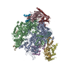
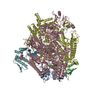
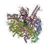
 マップデータ マップデータ | 3D reconstruction of the skeletal muscle dihydropyridin receptor (DHPR, L-type calcium channel) from single particle images. Low-pass filtered at 23A resolution. Ref.: J Mol Biol. 2003 Sep 5;332(1):171-82 If displayed with chimera (http://www.cgl.ucsf.edu/chimera/download) threshold level for published protein surface (white transparent) = 0.0626 threshold level for published core density (red) = 0.0763 | |||||||||
 試料 試料 |
| |||||||||
| 生物種 |  | |||||||||
| 手法 | 単粒子再構成法 / クライオ電子顕微鏡法 / ネガティブ染色法 / 解像度: 21.0 Å | |||||||||
 データ登録者 データ登録者 | Wolf M / Eberhart A / Glossmann H / Striessnig J / Grigorieff N | |||||||||
 引用 引用 |  ジャーナル: J Mol Biol / 年: 2003 ジャーナル: J Mol Biol / 年: 2003タイトル: Visualization of the domain structure of an L-type Ca2+ channel using electron cryo-microscopy. 著者: M Wolf / A Eberhart / H Glossmann / J Striessnig / N Grigorieff /  要旨: The three-dimensional structure of the skeletal muscle voltage-gated L-type calcium channel (Ca(v)1.1; dihydropyridine receptor, DHPR) was determined using electron cryo-microscopy and single- ...The three-dimensional structure of the skeletal muscle voltage-gated L-type calcium channel (Ca(v)1.1; dihydropyridine receptor, DHPR) was determined using electron cryo-microscopy and single-particle averaging. The structure shows a single channel complex with an approximate total molecular mass of 550 kDa, corresponding to the five known subunits of the DHPR, and bound detergent and lipid. Features visible in our structure together with antibody labeling of the beta and alpha(2) subunits allowed us to assign locations for four of the five subunits within the structure. The most striking feature of the structure is the extra-cellular alpha(2) subunit that protrudes from the membrane domain in close proximity to the alpha(1) subunit. The cytosolic beta subunit is located close to the membrane and adjacent to subunits alpha(1), gamma and delta. Our structure correlates well with the functional and biochemical data available for this channel and suggests a three-dimensional model for the excitation-contraction coupling complex consisting of DHPR tetrads and the calcium release channel. | |||||||||
| 履歴 |
|
- 構造の表示
構造の表示
| ムービー |
 ムービービューア ムービービューア |
|---|---|
| 構造ビューア | EMマップ:  SurfView SurfView Molmil Molmil Jmol/JSmol Jmol/JSmol |
| 添付画像 |
- ダウンロードとリンク
-EMDBアーカイブ
| マップデータ |  emd_1069.map.gz emd_1069.map.gz | 7.5 MB |  EMDBマップデータ形式 EMDBマップデータ形式 | |
|---|---|---|---|---|
| ヘッダ (付随情報) |  emd-1069-v30.xml emd-1069-v30.xml emd-1069.xml emd-1069.xml | 11.4 KB 11.4 KB | 表示 表示 |  EMDBヘッダ EMDBヘッダ |
| 画像 |  1069.gif 1069.gif | 42.2 KB | ||
| アーカイブディレクトリ |  http://ftp.pdbj.org/pub/emdb/structures/EMD-1069 http://ftp.pdbj.org/pub/emdb/structures/EMD-1069 ftp://ftp.pdbj.org/pub/emdb/structures/EMD-1069 ftp://ftp.pdbj.org/pub/emdb/structures/EMD-1069 | HTTPS FTP |
-検証レポート
| 文書・要旨 |  emd_1069_validation.pdf.gz emd_1069_validation.pdf.gz | 190.8 KB | 表示 |  EMDB検証レポート EMDB検証レポート |
|---|---|---|---|---|
| 文書・詳細版 |  emd_1069_full_validation.pdf.gz emd_1069_full_validation.pdf.gz | 189.9 KB | 表示 | |
| XML形式データ |  emd_1069_validation.xml.gz emd_1069_validation.xml.gz | 5.4 KB | 表示 | |
| アーカイブディレクトリ |  https://ftp.pdbj.org/pub/emdb/validation_reports/EMD-1069 https://ftp.pdbj.org/pub/emdb/validation_reports/EMD-1069 ftp://ftp.pdbj.org/pub/emdb/validation_reports/EMD-1069 ftp://ftp.pdbj.org/pub/emdb/validation_reports/EMD-1069 | HTTPS FTP |
-関連構造データ
- リンク
リンク
| EMDBのページ |  EMDB (EBI/PDBe) / EMDB (EBI/PDBe) /  EMDataResource EMDataResource |
|---|
- マップ
マップ
| ファイル |  ダウンロード / ファイル: emd_1069.map.gz / 形式: CCP4 / 大きさ: 7.8 MB / タイプ: IMAGE STORED AS FLOATING POINT NUMBER (4 BYTES) ダウンロード / ファイル: emd_1069.map.gz / 形式: CCP4 / 大きさ: 7.8 MB / タイプ: IMAGE STORED AS FLOATING POINT NUMBER (4 BYTES) | ||||||||||||||||||||||||||||||||||||||||||||||||||||||||||||||||||||
|---|---|---|---|---|---|---|---|---|---|---|---|---|---|---|---|---|---|---|---|---|---|---|---|---|---|---|---|---|---|---|---|---|---|---|---|---|---|---|---|---|---|---|---|---|---|---|---|---|---|---|---|---|---|---|---|---|---|---|---|---|---|---|---|---|---|---|---|---|---|
| 注釈 | 3D reconstruction of the skeletal muscle dihydropyridin receptor (DHPR, L-type calcium channel) from single particle images. Low-pass filtered at 23A resolution. Ref.: J Mol Biol. 2003 Sep 5;332(1):171-82 If displayed with chimera (http://www.cgl.ucsf.edu/chimera/download) threshold level for published protein surface (white transparent) = 0.0626 threshold level for published core density (red) = 0.0763 | ||||||||||||||||||||||||||||||||||||||||||||||||||||||||||||||||||||
| 投影像・断面図 | 画像のコントロール
画像は Spider により作成 | ||||||||||||||||||||||||||||||||||||||||||||||||||||||||||||||||||||
| ボクセルのサイズ | X=Y=Z: 3.39 Å | ||||||||||||||||||||||||||||||||||||||||||||||||||||||||||||||||||||
| 密度 |
| ||||||||||||||||||||||||||||||||||||||||||||||||||||||||||||||||||||
| 対称性 | 空間群: 1 | ||||||||||||||||||||||||||||||||||||||||||||||||||||||||||||||||||||
| 詳細 | EMDB XML:
CCP4マップ ヘッダ情報:
| ||||||||||||||||||||||||||||||||||||||||||||||||||||||||||||||||||||
-添付データ
- 試料の構成要素
-全体 : skeletal muscle dihydropyridine receptor
| 全体 | 名称: skeletal muscle dihydropyridine receptor |
|---|---|
| 要素 |
|
-超分子 #1000: skeletal muscle dihydropyridine receptor
| 超分子 | 名称: skeletal muscle dihydropyridine receptor / タイプ: sample / ID: 1000 / 詳細: channel complex is solubilized in Digitonin 集合状態: monodisperse channel-detergent complex consisting of 5 subunits Number unique components: 1 |
|---|---|
| 分子量 | 実験値: 800 KDa / 理論値: 550 KDa / 手法: gel filtration chromatography |
-分子 #1: DHPR with subunits alpha1, alpha2, beta, gamma, delta
| 分子 | 名称: DHPR with subunits alpha1, alpha2, beta, gamma, delta タイプ: protein_or_peptide / ID: 1 Name.synonym: DHPR, dihydropyridine receptor, CAv1.1, L type calcium channel 詳細: additional MW due to detergent and residual lipid; solubilized in Digitonin コピー数: 1 集合状態: monodisperse channel detergent complex with 5 subunits 組換発現: No |
|---|---|
| 由来(天然) | 生物種:  |
| 分子量 | 実験値: 550 KDa / 理論値: 800 KDa |
-実験情報
-構造解析
| 手法 | ネガティブ染色法, クライオ電子顕微鏡法 |
|---|---|
 解析 解析 | 単粒子再構成法 |
| 試料の集合状態 | particle |
- 試料調製
試料調製
| 濃度 | 0.35 mg/mL |
|---|---|
| 緩衝液 | pH: 7.4 詳細: 50mM Tris-HCl pH7.4, 150mM NaCl, 25uM CaCl2, 1mM Iodoacetamide, 0.1mM Benzamidine, 0.1mM PMSF, 0.1% (w/v) Digitonin. |
| 染色 | タイプ: NEGATIVE 詳細: 5ul sample on holey carbon grids (Quantifoil) were vitrified by plunging into liqid ethane after blotting with filter paper. |
| グリッド | 詳細: Quantifoil (R) holey carbon copper grids |
| 凍結 | 凍結剤: ETHANE / チャンバー内湿度: 80 % / チャンバー内温度: 20 K / 装置: HOMEMADE PLUNGER / 詳細: Vitrification instrument: custom plunger 手法: Blot 5ul sample on negative glow-discharged grids for 5-10sec with Whatman filter paper before plunging. |
- 電子顕微鏡法
| 顕微鏡 | FEI TECNAI F20 |
|---|---|
| 温度 | 最低: 90 K / 最高: 95 K / 平均: 90 K |
| アライメント法 | Legacy - 非点収差: obj lens astigmatism was corrected at 270,000x magnification |
| 日付 | 2002年2月5日 |
| 撮影 | カテゴリ: FILM / フィルム・検出器のモデル: KODAK SO-163 FILM / デジタル化 - スキャナー: ZEISS SCAI / デジタル化 - サンプリング間隔: 7 µm / 実像数: 180 / 平均電子線量: 9 e/Å2 詳細: full scan resolution was downsampled by 3x3 pixel averaging. Od range: 1 / ビット/ピクセル: 8 |
| Tilt angle min | 0 |
| Tilt angle max | 0 |
| 電子線 | 加速電圧: 200 kV / 電子線源:  FIELD EMISSION GUN FIELD EMISSION GUN |
| 電子光学系 | 照射モード: FLOOD BEAM / 撮影モード: BRIGHT FIELD / Cs: 2.0 mm / 最大 デフォーカス(公称値): 4.5 µm / 最小 デフォーカス(公称値): 3.0 µm / 倍率(公称値): 62000 |
| 試料ステージ | 試料ホルダー: Side entry liquid nitrogen-cooled cryo holder 試料ホルダーモデル: GATAN LIQUID NITROGEN |
| 実験機器 |  モデル: Tecnai F20 / 画像提供: FEI Company |
- 画像解析
画像解析
| CTF補正 | 詳細: per micrograph, CTFFIT3 |
|---|---|
| 最終 再構成 | 想定した対称性 - 点群: C1 (非対称) / アルゴリズム: OTHER / 解像度のタイプ: BY AUTHOR / 解像度: 21.0 Å / 解像度の算出法: FSC 0.5 CUT-OFF / ソフトウェア - 名称: MRC, IMAGIC, FREALIGN 詳細: FREALIGN does not use class averages but performs an orientation search of each individual particle incl. CTF correction. The final map was reconstructed from contributions of 14,056 ...詳細: FREALIGN does not use class averages but performs an orientation search of each individual particle incl. CTF correction. The final map was reconstructed from contributions of 14,056 particles and sharpened with a negative temperature factor of 500. The absolute handedness of the reconstruction was not determined. 使用した粒子像数: 14056 |
 ムービー
ムービー コントローラー


 Z (Sec.)
 Y (Row.)
 X (Col.)


