 Movie
Movie Controller

[English] 日本語
 Yorodumi
- EMDB-1062: Three-dimensional structure of C complex spliceosomes by electron... -
+ Open data

- Basic information
| Entry | Database: EMDB / ID: EMD-1062 | |||||||||
|---|---|---|---|---|---|---|---|---|---|---|
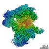
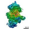
| Title | Three-dimensional structure of C complex spliceosomes by electron microscopy. | |||||||||
 Map data Map data | Map of C-complex of the human spliceosome. | |||||||||
 Sample Sample |
| |||||||||
| Biological species |   Homo sapiens (human) Homo sapiens (human) | |||||||||
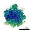
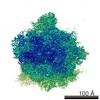
| Method | single particle reconstruction / cryo EM / negative staining / Resolution: 30.0 Å | |||||||||
 Authors Authors | Jurica MS / Sousa D / Moore MJ / Grigorieff N | |||||||||
 Citation Citation | Journal: Nat Struct Mol Biol / Year: 2004 Title: Three-dimensional structure of C complex spliceosomes by electron microscopy. Authors: Melissa S Jurica / Duncan Sousa / Melissa J Moore / Nikolaus Grigorieff /  Abstract: The spliceosome is a multimegadalton RNA-protein machine that removes noncoding sequences from nascent pre-mRNAs. Recruitment of the spliceosome to splice sites and subsequent splicing require a ...The spliceosome is a multimegadalton RNA-protein machine that removes noncoding sequences from nascent pre-mRNAs. Recruitment of the spliceosome to splice sites and subsequent splicing require a series of dynamic interactions among the spliceosome's component U snRNPs and many additional protein factors. These dynamics present several challenges for structural analyses, including purification of stable complexes to compositional homogeneity and assessment of conformational heterogeneity. We have isolated spliceosomes arrested before the second chemical step of splicing (C complex) in which U2, U5 and U6 snRNAs are stably associated. Using electron microscopy, we obtained images of C complex spliceosomes under cryogenic conditions and determined a three-dimensional structure of a core complex to a resolution of 30 A. The structure reveals a particle of dimensions 27 x 22 x 24 nm with a relatively open arrangement of three primary domains. | |||||||||
| History |
|
- Structure visualization
| Movie |
 Movie viewer Movie viewer |
|---|---|
| Structure viewer | EM map: SurfViewMolmilJmol/JSmol |
| Supplemental images |

- Downloads & links
-EMDB archive
| Map data | emd_1062.map.gz | 3.3 MB | EMDB map data format | |
|---|---|---|---|---|
| Header (meta data) | emd-1062-v30.xmlemd-1062.xml | 9.8 KB 9.8 KB | Display Display | EMDB header |
| Images |  1062.gif 1062.gif | 46.7 KB | ||
| Archive directory |  http://ftp.pdbj.org/pub/emdb/structures/EMD-1062ftp://ftp.pdbj.org/pub/emdb/structures/EMD-1062 http://ftp.pdbj.org/pub/emdb/structures/EMD-1062ftp://ftp.pdbj.org/pub/emdb/structures/EMD-1062 | HTTPS FTP |
-Related structure data
| Similar structure data |
|---|


-Links
| EMDB pages | EMDB (EBI/PDBe) / EMDataResource |
|---|
-Map
| File | Download / File: emd_1062.map.gz / Format: CCP4 / Size: 7.8 MB / Type: IMAGE STORED AS FLOATING POINT NUMBER (4 BYTES) | ||||||||||||||||||||||||||||||||||||||||||||||||||||||||||||||||||||
|---|---|---|---|---|---|---|---|---|---|---|---|---|---|---|---|---|---|---|---|---|---|---|---|---|---|---|---|---|---|---|---|---|---|---|---|---|---|---|---|---|---|---|---|---|---|---|---|---|---|---|---|---|---|---|---|---|---|---|---|---|---|---|---|---|---|---|---|---|---|
| Annotation | Map of C-complex of the human spliceosome. | ||||||||||||||||||||||||||||||||||||||||||||||||||||||||||||||||||||
| Voxel size | X=Y=Z: 4.6667 Å | ||||||||||||||||||||||||||||||||||||||||||||||||||||||||||||||||||||
| Density |
| ||||||||||||||||||||||||||||||||||||||||||||||||||||||||||||||||||||
| Symmetry | Space group: 1 | ||||||||||||||||||||||||||||||||||||||||||||||||||||||||||||||||||||
| Details | EMDB XML:
CCP4 map header:
| ||||||||||||||||||||||||||||||||||||||||||||||||||||||||||||||||||||
-Supplemental data
- Sample components
-Entire : C-complex of the human spliceosome
| Entire | Name: C-complex of the human spliceosome |
|---|---|
| Components |
|
-Supramolecule #1000: C-complex of the human spliceosome
| Supramolecule | Name: C-complex of the human spliceosome / type: sample / ID: 1000 / Number unique components: 1 |
|---|---|
| Molecular weight | Theoretical: 2.6 MDa Method: The molecular weight of core C-complex components were added together. The map becomes discontinous at either higher or lower weights. |
-Supramolecule #1: C-complex Spliceosome
| Supramolecule | Name: C-complex Spliceosome / type: organelle_or_cellular_component / ID: 1 / Name.synonym: Spliceosome / Number of copies: 1 / Oligomeric state: monomer / Recombinant expression: No |
|---|---|
| Source (natural) | Organism: Homo sapiens (human) / synonym: Human / Cell: HeLa / Organelle: Nucleus / Location in cell: Nucleus |
| Molecular weight | Experimental: 2.6 MDa |
-Experimental details
-Structure determination
| Method | negative staining, cryo EM |
|---|---|
 Processing Processing | single particle reconstruction |
| Aggregation state | particle |
-Sample preparation
| Buffer | pH: 7.5 Details: 150mM KCl, 5mM EDTA, 20mM Tris, 10mM maltose, and 5% glycerol |
|---|---|
| Staining | Type: NEGATIVE Details: Thin carbon is floated for 1 minute on the sample. The carbon is transferred onto a solution of 1% uranyl formate, sample side down, and then picked up with a holey carbon grid (Quantifoil). ...Details: Thin carbon is floated for 1 minute on the sample. The carbon is transferred onto a solution of 1% uranyl formate, sample side down, and then picked up with a holey carbon grid (Quantifoil). A second layer of thin carbon is sandwiched on top of the sample. |
| Grid | Details: 400 mesh copper grid |
| Vitrification | Cryogen name: NITROGEN / Instrument: HOMEMADE PLUNGER / Details: Vitrification instrument: human hand. none / Method: See experimental details. |
- Electron microscopy
| Microscope | FEI/PHILIPS CM12 |
|---|---|
| Electron beam | Acceleration voltage: 120 kV / Electron source: LAB6 |
| Electron optics | Calibrated magnification: 60000 / Illumination mode: FLOOD BEAM / Imaging mode: BRIGHT FIELDBright-field microscopy / Cs: 2 mm / Nominal defocus max: 20.0 µm / Nominal defocus min: 1.0 µm / Nominal magnification: 60000 |
| Sample stage | Specimen holder: Side entry liquid nitrogen-cooled cryo specimen holder. Specimen holder model: OTHER / Tilt angle min: 35 / Tilt angle max: 45 |
| Temperature | Min: 90 K / Max: 100 K / Average: 95 K |
| Alignment procedure | Legacy - Astigmatism: objective lens astigmatism was corrected at 200,000 times magnification |
| Details | MICROSCOPE Philips CM12 |
| Image recording | Category: FILM / Film or detector model: KODAK SO-163 FILM / Digitization - Scanner: ZEISS SCAI / Digitization - Sampling interval: 7 µm / Number real images: 72 / Average electron dose: 10 e/Å2 / Bits/pixel: 8 |
-Image processing
| CTF correction | Details: CTF correction of each particle |
|---|---|
| Final reconstruction | Applied symmetry - Point group: C1 (asymmetric) / Algorithm: OTHER / Resolution.type: BY AUTHOR / Resolution: 30.0 Å / Resolution method: FSC 0.5 CUT-OFF / Software - Name: Spider and Frealign Details: The final map was calculated from one dataset and filtered to 20 angstroms resolution. Number images used: 1834 |
| Details | Image tilt pairs were collected manually and processed using Spider and Frealign. |
