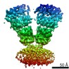
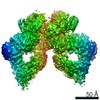
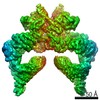
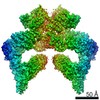
Journal: Nature / Year: 2020 Title: Structure of SAGA and mechanism of TBP deposition on gene promoters. Authors: Gabor Papai / Alexandre Frechard / Olga Kolesnikova / Corinne Crucifix / Patrick Schultz / Adam Ben-Shem / Abstract: SAGA (Spt-Ada-Gcn5-acetyltransferase) is a 19-subunit complex that stimulates transcription via two chromatin-modifying enzymatic modules and by delivering the TATA box binding protein (TBP) to ...SAGA (Spt-Ada-Gcn5-acetyltransferase) is a 19-subunit complex that stimulates transcription via two chromatin-modifying enzymatic modules and by delivering the TATA box binding protein (TBP) to nucleate the pre-initiation complex on DNA, a pivotal event in the expression of protein-encoding genes. Here we present the structure of yeast SAGA with bound TBP. The core of the complex is resolved at 3.5 Å resolution (0.143 Fourier shell correlation). The structure reveals the intricate network of interactions that coordinate the different functional domains of SAGA and resolves an octamer of histone-fold domains at the core of SAGA. This deformed octamer deviates considerably from the symmetrical analogue in the nucleosome and is precisely tuned to establish a peripheral site for TBP, where steric hindrance represses binding of spurious DNA. Complementary biochemical analysis points to a mechanism for TBP delivery and release from SAGA that requires transcription factor IIA and whose efficiency correlates with the affinity of DNA to TBP. We provide the foundations for understanding the specific delivery of TBP to gene promoters and the multiple roles of SAGA in regulating gene expression.
History
Deposition
Oct 31, 2019
-
Header (metadata) release
Jan 29, 2020
-
Map release
Jan 29, 2020
-
Update
Jun 30, 2021
-
Current status
Jun 30, 2021
Processing site: PDBe / Status: Released
-
Structure visualization
Movie
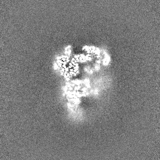
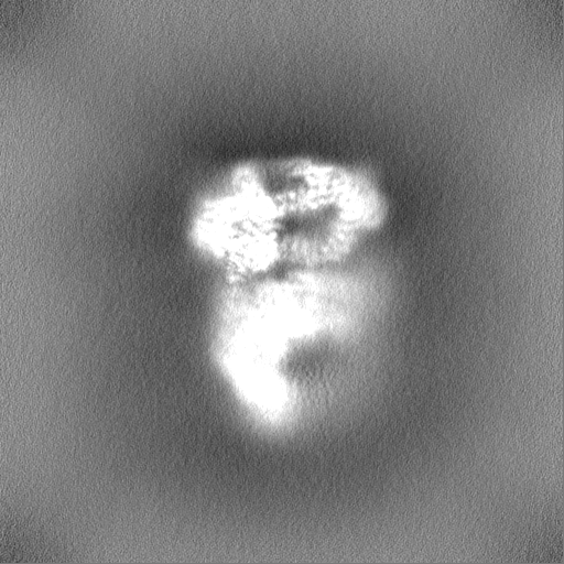
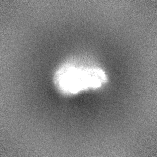
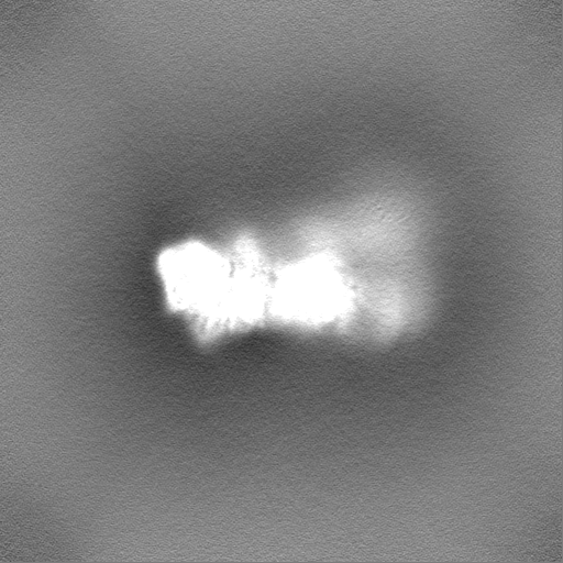
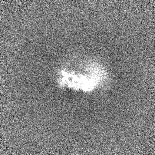
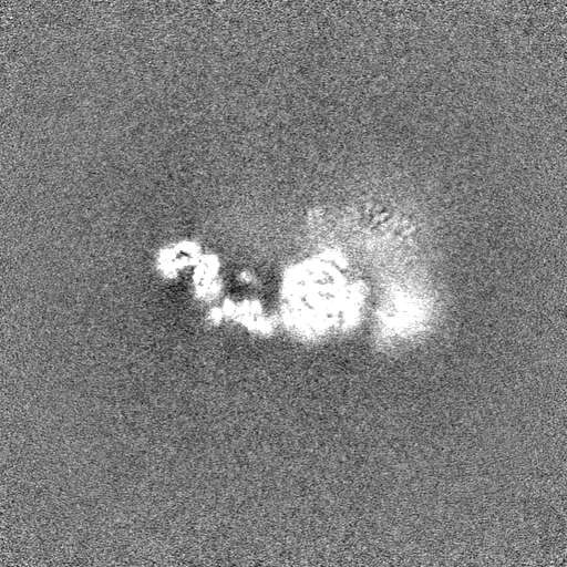
Surface view with section colored by density value
Macromolecule #5: Subunit of the SAGA and SAGA-like transcriptional regulatory comp...
Macromolecule
Name: Subunit of the SAGA and SAGA-like transcriptional regulatory complexes, interacts with Spt15p to act type: protein_or_peptide / ID: 5 / Number of copies: 1 / Enantiomer: LEVO
Macromolecule #6: Subunit of the SAGA transcriptional regulatory complex, involved ...
Macromolecule
Name: Subunit of the SAGA transcriptional regulatory complex, involved in proper assembly of the complex type: protein_or_peptide / ID: 6 / Number of copies: 1 / Enantiomer: LEVO
Macromolecule #11: Subunit (17 kDa) of TFIID and SAGA complexes, involved in RNA pol...
Macromolecule
Name: Subunit (17 kDa) of TFIID and SAGA complexes, involved in RNA polymerase II transcription initiation type: protein_or_peptide / ID: 11 / Number of copies: 1 / Enantiomer: LEVO
Film or detector model: GATAN K2 QUANTUM (4k x 4k) / Detector mode: SUPER-RESOLUTION / Average exposure time: 8.0 sec. / Average electron dose: 52.8 e/Å2
Experimental equipment
Model: Titan Krios / Image courtesy: FEI Company
-
Image processing
Particle selection
Number selected: 1068534
CTF correction
Software - Name: Warp
Initial angle assignment
Type: MAXIMUM LIKELIHOOD / Software - Name: cryoSPARC
Final angle assignment
Type: MAXIMUM LIKELIHOOD / Software - Name: cryoSPARC
Final reconstruction
Applied symmetry - Point group: C1 (asymmetric) / Algorithm: FOURIER SPACE / Resolution.type: BY AUTHOR / Resolution: 3.8 Å / Resolution method: FSC 0.143 CUT-OFF / Number images used: 354104
In the structure databanks used in Yorodumi, some data are registered as the other names, "COVID-19 virus" and "2019-nCoV". Here are the details of the virus and the list of structure data.
Jan 31, 2019. EMDB accession codes are about to change! (news from PDBe EMDB page)
EMDB accession codes are about to change! (news from PDBe EMDB page)
The allocation of 4 digits for EMDB accession codes will soon come to an end. Whilst these codes will remain in use, new EMDB accession codes will include an additional digit and will expand incrementally as the available range of codes is exhausted. The current 4-digit format prefixed with “EMD-” (i.e. EMD-XXXX) will advance to a 5-digit format (i.e. EMD-XXXXX), and so on. It is currently estimated that the 4-digit codes will be depleted around Spring 2019, at which point the 5-digit format will come into force.
The EM Navigator/Yorodumi systems omit the EMD- prefix.
Related info.:Q: What is EMD? / ID/Accession-code notation in Yorodumi/EM Navigator
Yorodumi is a browser for structure data from EMDB, PDB, SASBDB, etc.
This page is also the successor to EM Navigator detail page, and also detail information page/front-end page for Omokage search.
The word "yorodu" (or yorozu) is an old Japanese word meaning "ten thousand". "mi" (miru) is to see.
Related info.:EMDB / PDB / SASBDB / Comparison of 3 databanks / Yorodumi Search / Aug 31, 2016. New EM Navigator & Yorodumi / Yorodumi Papers / Jmol/JSmol / Function and homology information / Changes in new EM Navigator and Yorodumi
 Movie
Movie Controller


 Open data
 Basic information
 Map data
 Sample
 Function and homology information
 transcription initiation at RNA polymerase I promoter / RNA polymerase II core promoter sequence-specific DNA binding / RNA polymerase II preinitiation complex assembly / DNA-templated transcription initiation / chromatin organization / RNA polymerase II-specific DNA-binding transcription factor binding / transcription by RNA polymerase II /
  Komagataella phaffii GS115 (fungus) /
  Authors
 France, 3 items
  Citation
 Structure visualization
 Downloads & links
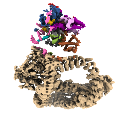 emd_10438.png
 http://ftp.pdbj.org/pub/emdb/structures/EMD-10438


 Z
Z Y
Y X
X


 Sample components
 Processing
 Electron microscopy
