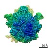
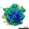
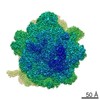
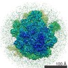
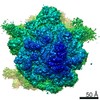
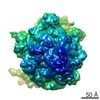
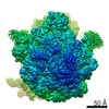
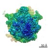
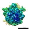
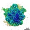
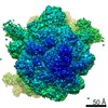
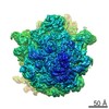
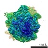
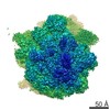
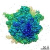
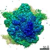
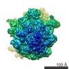
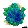
- EMDB-1003: Solution structure of the E. coli 70S ribosome at 11.5 A resolution. -
+
データを開く
IDまたはキーワード:
読み込み中...
-
基本情報
登録情報
データベース: EMDB / ID: EMD-1003
タイトル
Solution structure of the E. coli 70S ribosome at 11.5 A resolution.
マップデータ
E. coli 70S Ribosome
試料
試料: FMet-tRNAMet 70S Ribosome from E.coli
複合体: 70S ribosome Escherichia coli
RNA: fMet-tRNA
RNA: MF-mRNA
機能・相同性
機能・相同性情報
ribosomal small subunit biogenesis / cytosolic small ribosomal subunit / large ribosomal subunit rRNA binding / large ribosomal subunit / regulation of translation / small ribosomal subunit / cytosolic large ribosomal subunit / tRNA binding / rRNA binding / リボソーム ...ribosomal small subunit biogenesis / cytosolic small ribosomal subunit / large ribosomal subunit rRNA binding / large ribosomal subunit / regulation of translation / small ribosomal subunit / cytosolic large ribosomal subunit / tRNA binding / rRNA binding / リボソーム / structural constituent of ribosome / 翻訳 (生物学) / ribonucleoprotein complex / response to antibiotic / 細胞質 類似検索 - 分子機能
Ribosomal protein L1, bacterial-type / Ribosomal protein L1, conserved site / Ribosomal protein L1 / Ribosomal protein L1 signature. / Ribosomal protein L1, 3-layer alpha/beta-sandwich / Ribosomal protein L1-like / Ribosomal protein L1/ribosomal biogenesis protein / Ribosomal protein L1p/L10e family / Ribosomal protein L11, bacterial-type / Ribosomal protein L11, conserved site ...Ribosomal protein L1, bacterial-type / Ribosomal protein L1, conserved site / Ribosomal protein L1 / Ribosomal protein L1 signature. / Ribosomal protein L1, 3-layer alpha/beta-sandwich / Ribosomal protein L1-like / Ribosomal protein L1/ribosomal biogenesis protein / Ribosomal protein L1p/L10e family / Ribosomal protein L11, bacterial-type / Ribosomal protein L11, conserved site / Ribosomal protein L11 signature. / Ribosomal protein L6, conserved site / Ribosomal protein L6 signature 1. / Ribosomal protein L11, N-terminal / Ribosomal protein L11/L12 / Ribosomal protein L11, C-terminal / Ribosomal protein L11, C-terminal domain superfamily / Ribosomal protein L11/L12, N-terminal domain superfamily / Ribosomal protein L11/L12 / Ribosomal protein L11, N-terminal domain / Ribosomal protein L11, RNA binding domain / Ribosomal protein L6, bacterial-type / Ribosomal protein S6, conserved site / Ribosomal protein S6 signature. / Ribosomal protein S7, bacterial/organellar-type / Ribosomal protein S4, bacterial-type / 30S ribosomal protein S17 / Ribosomal protein S5, bacterial-type / Ribosomal protein S6, plastid/chloroplast / Ribosomal protein S15, bacterial-type / Ribosomal protein S6 / Ribosomal protein S6 / Ribosomal protein S6 superfamily / Translation elongation factor EF1B/ribosomal protein S6 / : / Ribosomal protein S7, conserved site / Ribosomal protein S17, conserved site / Ribosomal protein S4/S9 N-terminal domain / Ribosomal protein S4/S9, N-terminal / Ribosomal protein S4, conserved site / Ribosomal protein S4/S9 N-terminal domain / Ribosomal protein S4/S9 / Ribosomal protein S8 / Ribosomal protein S8 superfamily / Ribosomal protein S5, N-terminal, conserved site / Ribosomal protein S5 signature. / Ribosomal protein L6, alpha-beta domain / Ribosomal protein L6 / Ribosomal protein L6, alpha-beta domain superfamily / Ribosomal protein S7 signature. / Ribosomal protein L6 / S5 double stranded RNA-binding domain profile. / Ribosomal protein S5 / Ribosomal protein S5, N-terminal / Ribosomal protein S8 / Ribosomal protein S5, N-terminal domain / Ribosomal protein S5, C-terminal / Ribosomal protein S17 signature. / S4 RNA-binding domain / Ribosomal protein S5, C-terminal domain / RNA-binding S4 domain / Ribosomal protein S5/S7 / Ribosomal protein S7 domain / Ribosomal protein S7 domain superfamily / RNA-binding S4 domain superfamily / Ribosomal protein S8 signature. / S4 domain / Ribosomal protein S17/S11 / Ribosomal protein S4 signature. / Ribosomal_S15 / Ribosomal protein S15 signature. / Ribosomal protein S15 / Ribosomal protein S7p/S5e / S4 RNA-binding domain profile. / Ribosomal protein S17 / Ribosomal protein S15 / S15/NS1, RNA-binding / Ribosomal protein S5 domain 2-type fold, subgroup / Ribosomal protein S5 domain 2-type fold / Nucleic acid-binding, OB-fold 類似検索 - ドメイン・相同性
Small ribosomal subunit protein uS5 / Large ribosomal subunit protein uL6 / Small ribosomal subunit protein uS8 / Small ribosomal subunit protein uS7 / Small ribosomal subunit protein uS4 / Small ribosomal subunit protein uS5 / Small ribosomal subunit protein bS6 / Small ribosomal subunit protein uS17 / Large ribosomal subunit protein uL1 / Large ribosomal subunit protein uL11 ...Small ribosomal subunit protein uS5 / Large ribosomal subunit protein uL6 / Small ribosomal subunit protein uS8 / Small ribosomal subunit protein uS7 / Small ribosomal subunit protein uS4 / Small ribosomal subunit protein uS5 / Small ribosomal subunit protein bS6 / Small ribosomal subunit protein uS17 / Large ribosomal subunit protein uL1 / Large ribosomal subunit protein uL11 / Small ribosomal subunit protein uS15 / Small ribosomal subunit protein uS4 / Large ribosomal subunit protein uL6 / Small ribosomal subunit protein uS15 / Large ribosomal subunit protein uL1 / Small ribosomal subunit protein bS6 類似検索 - 構成要素
ジャーナル: Cell / 年: 2000 タイトル: Solution structure of the E. coli 70S ribosome at 11.5 A resolution. 著者: I S Gabashvili / R K Agrawal / C M Spahn / R A Grassucci / D I Svergun / J Frank / P Penczek / 要旨: Over 73,000 projections of the E. coli ribosome bound with formyl-methionyl initiator tRNAf(Met) were used to obtain an 11.5 A cryo-electron microscopy map of the complex. This map allows ...Over 73,000 projections of the E. coli ribosome bound with formyl-methionyl initiator tRNAf(Met) were used to obtain an 11.5 A cryo-electron microscopy map of the complex. This map allows identification of RNA helices, peripheral proteins, and intersubunit bridges. Comparison of double-stranded RNA regions and positions of proteins identified in both cryo-EM and X-ray maps indicates good overall agreement but points to rearrangements of ribosomal components required for the subunit association. Fitting of known components of the 50S stalk base region into the map defines the architecture of the GTPase-associated center and reveals a major change in the orientation of the alpha-sarcin-ricin loop. Analysis of the bridging connections between the subunits provides insight into the dynamic signaling mechanism between the ribosomal subunits.
ダウンロード / ファイル: emd_1003.map.gz / 形式: CCP4 / 大きさ: 7.3 MB / タイプ: IMAGE STORED AS FLOATING POINT NUMBER (4 BYTES)
注釈
E. coli 70S Ribosome
ボクセルのサイズ
X=Y=Z: 2.93 Å
密度
表面レベル
1: 82.799999999999997 / ムービー #1: 50
最小 - 最大
-141.974999999999994 - 339.987000000000023
平均 (標準偏差)
1.85585 (±38.046700000000001)
対称性
空間群: 1
詳細
EMDB XML:
マップ形状
Axis order
X
Y
Z
Origin
-62
-62
-62
サイズ
125
125
125
Spacing
125
125
125
セル
A=B=C: 366.25 Å α=β=γ: 90 °
CCP4マップ ヘッダ情報:
mode
Image stored as Reals
Å/pix. X/Y/Z
2.93
2.93
2.93
M x/y/z
125
125
125
origin x/y/z
0.000
0.000
0.000
length x/y/z
366.250
366.250
366.250
α/β/γ
90.000
90.000
90.000
MAP C/R/S
1
2
3
start NC/NR/NS
-62
-62
-62
NC/NR/NS
125
125
125
D min/max/mean
-141.975
339.987
1.856
-
添付データ
-
試料の構成要素
-
全体 : FMet-tRNAMet 70S Ribosome from E.coli
全体
名称: FMet-tRNAMet 70S Ribosome from E.coli
要素
試料: FMet-tRNAMet 70S Ribosome from E.coli
複合体: 70S ribosome Escherichia coli
RNA: fMet-tRNA
RNA: MF-mRNA
-
超分子 #1000: FMet-tRNAMet 70S Ribosome from E.coli
超分子
名称: FMet-tRNAMet 70S Ribosome from E.coli / タイプ: sample / ID: 1000 詳細: preparation and buffer conditions are as described in Malhotra et al., J. Mol. Biol. (1998) 280, 103-116 Number unique components: 3
想定した対称性 - 点群: C1 (非対称) / アルゴリズム: OTHER / 解像度のタイプ: BY AUTHOR / 解像度: 11.5 Å / 解像度の算出法: FSC 0.5 CUT-OFF / ソフトウェア - 名称: SPIDER/WEB / 使用した粒子像数: 73523
-
原子モデル構築 1
ソフトウェア
名称: O
詳細
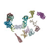
manual fitting using O Mol_Id: 1; Molecule: S4 Ribosomal Protein; Chain: A; Other_Details: Modeled By Analogous Protein Of B. Stearothermophilus Taken From Pdb Entry 1C06 Mol_Id: 2; Molecule: S5 Ribosomal Protein; Chain: B; Other_Details: Modeled By Analogous Protein Of B. Stearothermophilus Taken From Pdb Entry 1Pkp Mol_Id: 3; Molecule: S6 Ribosomal Protein; Chain: C; Other_Details: Modeled By Analogous Protein Of T. Thermophilus Taken From Pdb Entry 1Ris Mol_Id: 4; Molecule: S7 Ribosomal Protein; Chain: D; Other_Details: Modeled By Analogous Protein Of T. Thermophilus Taken From Pdb Entry 1Rss Mol_Id: 5; Molecule: S8 Ribosomal Protein; Chain: E; Other_Details: Modeled By Analogous Protein Of T. Thermophilus Taken From Pdb Entry 1An7 Mol_Id: 6; Molecule: S15 Ribosomal Protein; Chain: F; Other_Details: Modeled By Analogous Protein Of B. Stearothermophilus Taken From Pdb Entry 1A32 Mol_Id: 7; Molecule: S17 Ribosomal Protein; Chain: G; Other_Details: Modeled By Analogous Protein Of B. Stearothermophilus Taken From Pdb Entries 1Rip and 1Qd7 Mol_Id: 8; Molecule: S20 Ribosomal Protein; Chain: H; Other_Details: Modeled By Analogous Protein Of T. Thermophilus Taken From Pdb Entry 1Qd7 Mol_Id: 9; Molecule: Ribosomal Protein L1; Chain: N; Other_Details: Modeled By Analogous Protein Of T. Thermophilus Taken From Pdb Entry 1Ad2 Mol_Id: 10; Molecule: Ribosomal Protein L6; Chain: J; Mutation: Yes; Other_Details: Modeled By Analogous Protein Of T. Stearothermophilus Taken From Pdb Entry 1Rl6 Mol_Id: 11; Molecule: Ribosomal Protein L11; Chain: K; Other_Details: Modeled By Analogous Protein Of T. Maritima Taken From Pdb Entry 1Mms Mol_Id: 12; Molecule: Fragment Of 16S Rrna Helix 23; Chain: I; Fragment: Residues 673-713; Other_Details: Modeled As Analogous Fragment Of T. Thermophilus Taken From Pdb Entry 1Qd7 Mol_Id: 13; Molecule: Fragment Of 23S Rrna; Chain: L; Fragment: Residues 1051-1108; Other_Details: T. Maritima RNA Sequence and Model Taken From Pdb Entry 1Mms Mol_Id: 14; Molecule: Helix 95 Of 23S Rrna; Chain: M; Other_Details: E. Coli RNA Sequence and Model Taken From Pdb Entry 480D Mol_Id: 15; Molecule: Formyl-Methionyl-tRNA; Chain: O; Synonym: Fmet-tRNA; Other_Details: E. Coli Fmet-tRNA Sequence and Model Taken From Pdb Entry 2Fmt
精密化
プロトコル: RIGID BODY FIT
得られたモデル
PDB-1eg0: FITTING OF COMPONENTS WITH KNOWN STRUCTURE INTO AN 11.5 A CRYO-EM MAP OF THE E.COLI 70S RIBOSOME
 ムービー
ムービー コントローラー


 データを開く
 基本情報
基本情報 マップデータ
 試料
試料 機能・相同性情報
 ribosomal small subunit biogenesis / cytosolic small ribosomal subunit / large ribosomal subunit rRNA binding / large ribosomal subunit /
 データ登録者
 引用
引用
 構造の表示
構造の表示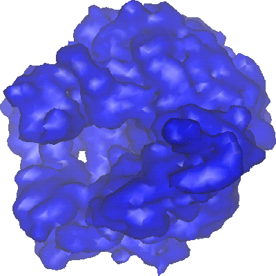
 ダウンロードとリンク
 1003.gif
 http://ftp.pdbj.org/pub/emdb/structures/EMD-1003
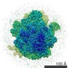


 試料の構成要素
 解析
解析 電子顕微鏡法