Loading
PDBj
MenuPDBj@FacebookPDBj@TwitterPDBj@YouTubewwPDB FoundationwwPDB
RCSB PDBPDBeBMRBAdv. SearchSearch help
Search by PDB author
1DT7
DownloadVisualize
BU of 1dt7 by Molmil
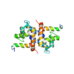
SOLUTION STRUCTURE OF THE C-TERMINAL NEGATIVE REGULATORY DOMAIN OF P53 IN A COMPLEX WITH CA2+-BOUND S100B(BB)
Descriptor: CALCIUM ION, CELLULAR TUMOR ANTIGEN P53, S100 CALCIUM-BINDING PROTEIN
Authors:Rustandi, R.R, Baldisseri, D.M, Weber, D.J.
Deposit date:2000-01-11
Release date:2000-07-26
Last modified:2024-04-10
Method:SOLUTION NMR
Cite:Structure of the negative regulatory domain of p53 bound to S100B(betabeta).
Nat.Struct.Biol., 7, 2000
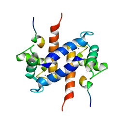
1K2H
DownloadVisualize
BU of 1k2h by Molmil
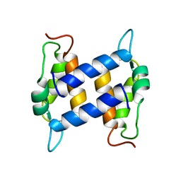
Three-dimensional Solution Structure of apo-S100A1.
Descriptor: S-100 protein, alpha chain
Authors:Rustandi, R.R, Baldisseri, D.M, Inman, K.G, Nizner, P, Hamilton, S.M, Landar, A, Landar, A, Zimmer, D.B, Weber, D.J.
Deposit date:2001-09-27
Release date:2002-02-13
Last modified:2017-02-01
Method:SOLUTION NMR
Cite:Three-dimensional solution structure of the calcium-signaling protein apo-S100A1 as determined by NMR.
Biochemistry, 41, 2002
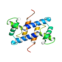
1MWN
DownloadVisualize
BU of 1mwn by Molmil
Solution NMR structure of S100B bound to the high-affinity target peptide TRTK-12
Descriptor: CALCIUM ION, F-actin capping protein alpha-1 subunit, S-100 protein, ...
Authors:Inman, K.G, Yang, R, Rustandi, R.R, Miller, K.E, Baldisseri, D.M, Weber, D.J.
Deposit date:2002-09-30
Release date:2002-12-18
Last modified:2022-02-23
Method:SOLUTION NMR
Cite:Solution NMR structure of S100B bound to the high-affinity target peptide TRTK-12
J.Mol.Biol., 324, 2002
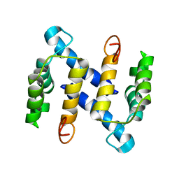
1M31
DownloadVisualize
BU of 1m31 by Molmil
Three-Dimensional Solution Structure of Apo-Mts1
Descriptor: Placental calcium-binding protein
Authors:Vallely, K.M, Rustandi, R.R, Ellis, K.C, Varlamova, O, Bresnick, A.R, Weber, D.J.
Deposit date:2002-06-26
Release date:2002-10-30
Last modified:2022-02-23
Method:SOLUTION NMR
Cite:Solution structure of human Mts1 (S100A4) as determined by NMR spectroscopy.
Biochemistry, 41, 2002
1QLK
DownloadVisualize
BU of 1qlk by Molmil
SOLUTION STRUCTURE OF CA(2+)-LOADED RAT S100B (BETABETA) NMR, 20 STRUCTURES
Descriptor: CALCIUM ION, S-100 PROTEIN
Authors:Drohat, A.C, Baldisseri, D.M, Rustandi, R.R, Weber, D.J.
Deposit date:1997-09-26
Release date:1998-11-11
Last modified:2022-03-02
Method:SOLUTION NMR
Cite:Solution structure of calcium-bound rat S100B(betabeta) as determined by nuclear magnetic resonance spectroscopy,.
Biochemistry, 37, 1998
8EKL
DownloadVisualize
BU of 8ekl by Molmil
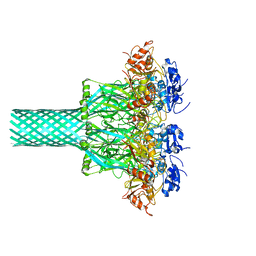
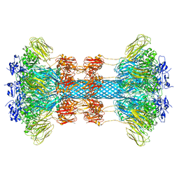
Clostridioides difficile binary toxin translocase CDTb wild-type after calcium depletion from receptor binding domain 1 (RBD1) - Class 1
Descriptor: ADP-ribosyltransferase binding component, CALCIUM ION
Authors:Abeyawardhane, D.L, Pozharski, E.
Deposit date:2022-09-21
Release date:2023-09-27
Method:ELECTRON MICROSCOPY (3.06 Å)
Cite:Calcium-mediated Pore Formation of Clostridioides difficile Binary Toxin
To Be Published
8EKM
DownloadVisualize
BU of 8ekm by Molmil
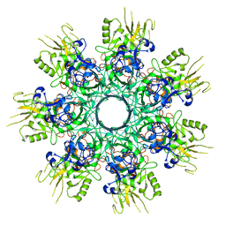
Clostridioides difficile binary toxin translocase CDTb double mutant - D623A D734A
Descriptor: ADP-ribosyltransferase binding component, CALCIUM ION
Authors:Abeyawardhane, D.L, Pozharski, E.
Deposit date:2022-09-21
Release date:2023-09-27
Method:ELECTRON MICROSCOPY (3.56 Å)
Cite:Calcium-mediated Pore Formation of Clostridioides difficile Binary Toxin
To Be Published
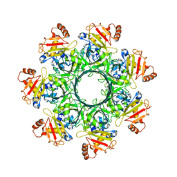
8EKK
DownloadVisualize
BU of 8ekk by Molmil
Clostridioides difficile binary toxin translocase CDTb wild-type after calcium depletion from receptor binding domain 1 (RBD1) - Class 2
Descriptor: ADP-ribosyltransferase binding component, CALCIUM ION
Authors:Abeyawardhane, D.L, Pozharski, E.
Deposit date:2022-09-21
Release date:2023-09-27
Method:ELECTRON MICROSCOPY (3.28 Å)
Cite:Calcium-mediated Pore Formation of Clostridioides difficile Binary Toxin
To Be Published
6UWI
DownloadVisualize
BU of 6uwi by Molmil
Crystal structure of the Clostridium difficile translocase CDTb
Descriptor: ADP-ribosyltransferase binding component, CALCIUM ION
Authors:Pozharski, E.
Deposit date:2019-11-05
Release date:2020-01-22
Last modified:2024-04-03
Method:X-RAY DIFFRACTION (3.7 Å)
Cite:Structure of the cell-binding component of theClostridium difficilebinary toxin reveals a di-heptamer macromolecular assembly.
Proc.Natl.Acad.Sci.USA, 117, 2020
6UWT
DownloadVisualize
BU of 6uwt by Molmil
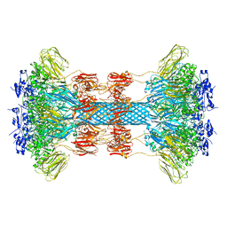
Clostridium difficile binary toxin translocase CDTb tetradecamer in symmetric conformation
Descriptor: ADP-ribosyltransferase binding component, CALCIUM ION
Authors:Xu, X, Pozharski, E, des Georges, A.
Deposit date:2019-11-05
Release date:2020-01-22
Last modified:2024-03-06
Method:ELECTRON MICROSCOPY (3.1 Å)
Cite:Structure of the cell-binding component of theClostridium difficilebinary toxin reveals a di-heptamer macromolecular assembly.
Proc.Natl.Acad.Sci.USA, 117, 2020
6UWR
DownloadVisualize
BU of 6uwr by Molmil
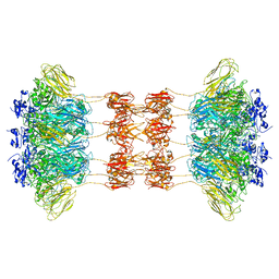
Clostridium difficile binary toxin translocase CDTb in asymmetric tetradecamer conformation
Descriptor: ADP-ribosyltransferase binding component, CALCIUM ION
Authors:Xu, X, Pozharski, E, des Georges, A.
Deposit date:2019-11-05
Release date:2020-01-22
Last modified:2024-03-06
Method:ELECTRON MICROSCOPY (2.8 Å)
Cite:Structure of the cell-binding component of theClostridium difficilebinary toxin reveals a di-heptamer macromolecular assembly.
Proc.Natl.Acad.Sci.USA, 117, 2020
6UWO
DownloadVisualize
BU of 6uwo by Molmil
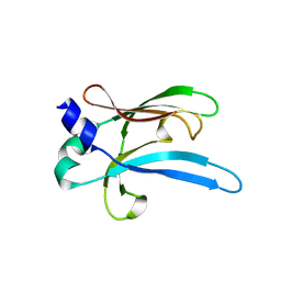
Crystal structure of receptor binding domain 2 from Clostridium difficile translocase CDTb
Descriptor: ADP-ribosyltransferase binding component
Authors:Pozharski, E.
Deposit date:2019-11-05
Release date:2020-01-22
Last modified:2024-03-06
Method:X-RAY DIFFRACTION (2.3 Å)
Cite:Structure of the cell-binding component of theClostridium difficilebinary toxin reveals a di-heptamer macromolecular assembly.
Proc.Natl.Acad.Sci.USA, 117, 2020

218853

PDB entries from 2024-04-24

PDB statisticsPDBj update infoContact PDBjnumon