Loading
PDBj
MenuPDBj@FacebookPDBj@TwitterPDBj@YouTubewwPDB FoundationwwPDB
RCSB PDBPDBeBMRBAdv. SearchSearch help
Search by PDB author
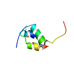
1JCO
DownloadVisualize
BU of 1jco by Molmil
Solution structure of the monomeric [Thr(B27)->Pro,Pro(B28)->Thr] insulin mutant (PT insulin)
Descriptor: Insulin A chain, Insulin B chain
Authors:Keller, D, Clausen, R, Josefsen, K, Led, J.J.
Deposit date:2001-06-11
Release date:2001-10-03
Last modified:2021-10-27
Method:SOLUTION NMR
Cite:Flexibility and bioactivity of insulin: an NMR investigation of the solution structure and folding of an unusually flexible human insulin mutant with increased biological activity.
Biochemistry, 40, 2001
1D0R
DownloadVisualize
BU of 1d0r by Molmil
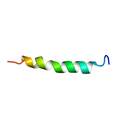
SOLUTION STRUCTURE OF GLUCAGON-LIKE PEPTIDE-1-(7-36)-AMIDE IN TRIFLUOROETHANOL/WATER
Descriptor: GLUCAGON-LIKE PEPTIDE-1-(7-36)-AMIDE
Authors:Chang, X, Keller, D, Bjorn, S, Led, J.J.
Deposit date:1999-09-14
Release date:2002-10-16
Last modified:2024-04-10
Method:SOLUTION NMR
Cite:Structure and Folding of Glucagon-like Peptide-1-(7-36)-amide in Trifluoroethanol Studied by NMR
MAGN.RESON.CHEM., 39, 2001
3Q0Y
DownloadVisualize
BU of 3q0y by Molmil
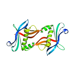
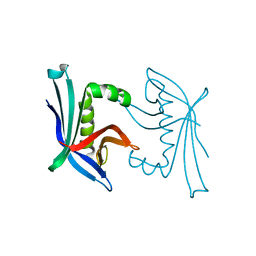
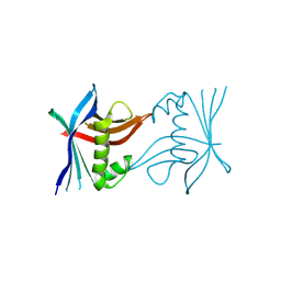
N-terminal domain of C. reinhardtii SAS-6 homolog Bld12p
Descriptor: Centriole protein
Authors:Kitagawa, D, Vakonakis, I, Olieric, N, Hilbert, M, Keller, D, Olieric, V, Bortfeld, M, Erat, M.C, Flueckiger, I, Goenczy, P, Steinmetz, M.O.
Deposit date:2010-12-16
Release date:2011-02-09
Last modified:2024-02-21
Method:X-RAY DIFFRACTION (2.1 Å)
Cite:Structural basis of the 9-fold symmetry of centrioles.
Cell(Cambridge,Mass.), 144, 2011
3Q0X
DownloadVisualize
BU of 3q0x by Molmil
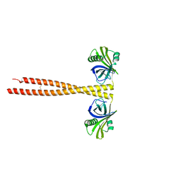
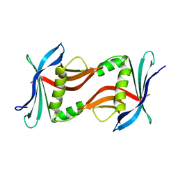
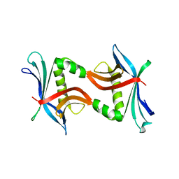
N-terminal coiled-coil dimer domain of C. reinhardtii SAS-6 homolog Bld12p
Descriptor: Centriole protein
Authors:Kitagawa, D, Vakonakis, I, Olieric, N, Hilbert, M, Keller, D, Olieric, V, Bortfeld, M, Erat, M.C, Flueckiger, I, Goenczy, P, Steinmetz, M.O.
Deposit date:2010-12-16
Release date:2011-02-09
Last modified:2011-07-13
Method:X-RAY DIFFRACTION (3.02 Å)
Cite:Structural basis of the 9-fold symmetry of centrioles.
Cell(Cambridge,Mass.), 144, 2011
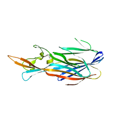
1T5R
DownloadVisualize
BU of 1t5r by Molmil
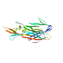
STRUCTURE OF THE PANTON-VALENTINE LEUCOCIDIN S COMPONENT FROM STAPHYLOCOCCUS AUREUS
Descriptor: LukS-PV
Authors:Guillet, V, Roblin, P, Keller, D, Prevost, G, Mourey, L.
Deposit date:2004-05-05
Release date:2004-08-24
Last modified:2023-08-23
Method:X-RAY DIFFRACTION (2 Å)
Cite:Crystal structure of leucotoxin S component: new insight into the Staphylococcal beta-barrel pore-forming toxins.
J.Biol.Chem., 279, 2004
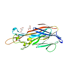
3PYI
DownloadVisualize
BU of 3pyi by Molmil
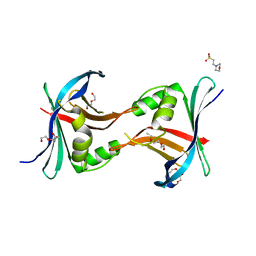
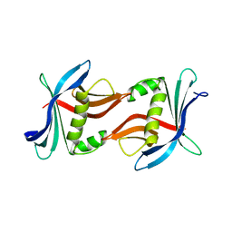
Structure of the N-terminal domain of C. elegans SAS-6
Descriptor: 2-(N-MORPHOLINO)-ETHANESULFONIC ACID, Spindle assembly abnormal protein 6, TETRAETHYLENE GLYCOL
Authors:Vakonakis, I, Steinmetz, M.O.
Deposit date:2010-12-13
Release date:2011-02-09
Last modified:2024-02-21
Method:X-RAY DIFFRACTION (2.104 Å)
Cite:Structural basis of the 9-fold symmetry of centrioles.
Cell(Cambridge,Mass.), 144, 2011
4TO7
DownloadVisualize
BU of 4to7 by Molmil
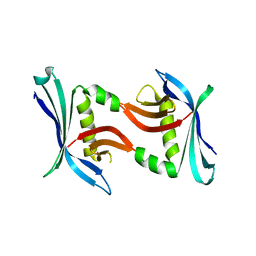
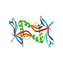
N-Terminal domain of C. Reinhardtii SAS-6 homolog bld12p Q93E F145W K146R (NN24)
Descriptor: Centriole protein
Authors:Hilbert, M, Kraatz, S.H.W.
Deposit date:2014-06-05
Release date:2015-06-17
Last modified:2023-12-20
Method:X-RAY DIFFRACTION (1.252 Å)
Cite:SAS-6 engineering reveals interdependence between cartwheel and microtubules in determining centriole architecture.
Nat.Cell Biol., 18, 2016
4TQ7
DownloadVisualize
BU of 4tq7 by Molmil
N-terminal domain of C. Reinhardtii SAS-6 homolog bld12p Q93E (NN10)
Descriptor: Centriole protein
Authors:Hilbert, M, Kraatz, S.H.W.
Deposit date:2014-06-10
Release date:2015-06-24
Last modified:2023-12-20
Method:X-RAY DIFFRACTION (2.643 Å)
Cite:SAS-6 engineering reveals interdependence between cartwheel and microtubules in determining centriole architecture.
Nat.Cell Biol., 18, 2016
4TTZ
DownloadVisualize
BU of 4ttz by Molmil
N-terminal domain of C. Reinhardtii SAS-6 homolog bld12p variant G94E F145W Q147K (NN26)
Descriptor: Centriole protein
Authors:Hilbert, M, Kraatz, S.H.W.
Deposit date:2014-06-23
Release date:2015-07-01
Last modified:2023-12-20
Method:X-RAY DIFFRACTION (2.5 Å)
Cite:SAS-6 engineering reveals interdependence between cartwheel and microtubules in determining centriole architecture.
Nat.Cell Biol., 18, 2016
4TTW
DownloadVisualize
BU of 4ttw by Molmil
N-terminal domain of C. Reinhardtii SAS-6 homolog bld12p K105C F145C (NN18)
Descriptor: Centriole protein
Authors:Hilbert, M, Kraatz, S.H.W.
Deposit date:2014-06-23
Release date:2015-07-01
Last modified:2023-12-20
Method:X-RAY DIFFRACTION (1.2 Å)
Cite:SAS-6 engineering reveals interdependence between cartwheel and microtubules in determining centriole architecture.
Nat.Cell Biol., 18, 2016
4TPZ
DownloadVisualize
BU of 4tpz by Molmil
N-terminal domain of C. Reinhardtii SAS-6 homolog bld12p F145W (NN2)
Descriptor: Centriole protein
Authors:Hilbert, M, Kraatz, S.H.W.
Deposit date:2014-06-10
Release date:2015-06-24
Last modified:2016-05-11
Method:X-RAY DIFFRACTION (1.8 Å)
Cite:SAS-6 engineering reveals interdependence between cartwheel and microtubules in determining centriole architecture.
Nat.Cell Biol., 18, 2016
4TTX
DownloadVisualize
BU of 4ttx by Molmil
N-terminal domain of C. Reinhardtii SAS-6 homolog bld12p variant G94C K146C (NN19)
Descriptor: Centriole protein
Authors:Hilbert, M, Kraatz, S.H.W.
Deposit date:2014-06-23
Release date:2015-07-01
Last modified:2023-12-20
Method:X-RAY DIFFRACTION (2.498 Å)
Cite:SAS-6 engineering reveals interdependence between cartwheel and microtubules in determining centriole architecture.
Nat.Cell Biol., 18, 2016
4U2J
DownloadVisualize
BU of 4u2j by Molmil
N-terminal domain of C. Reinhardtii SAS-6 homolog bld12p variant Q93E F145W (NN27)
Descriptor: Centriole protein
Authors:Hilbert, M, Kraatz, S.H.W.
Deposit date:2014-07-17
Release date:2015-07-29
Last modified:2016-05-11
Method:X-RAY DIFFRACTION (2 Å)
Cite:SAS-6 engineering reveals interdependence between cartwheel and microtubules in determining centriole architecture.
Nat.Cell Biol., 18, 2016
2QK7
DownloadVisualize
BU of 2qk7 by Molmil
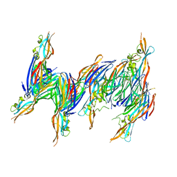
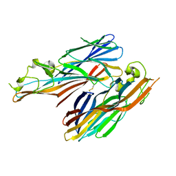
A covalent S-F heterodimer of staphylococcal gamma-hemolysin
Descriptor: Gamma-hemolysin component A, Gamma-hemolysin component B
Authors:Roblin, P, Guillet, V, Maveyraud, L, Mourey, L.
Deposit date:2007-07-10
Release date:2008-02-19
Last modified:2023-08-30
Method:X-RAY DIFFRACTION (2.4 Å)
Cite:A covalent S-F heterodimer of leucotoxin reveals molecular plasticity of beta-barrel pore-forming toxins.
Proteins, 71, 2008
4IYC
DownloadVisualize
BU of 4iyc by Molmil
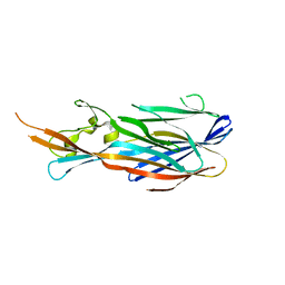
Structure of the T244A mutant of the PANTON-VALENTINE LEUCOCIDIN component from STAPHYLOCOCCUS AUREUS
Descriptor: LukS-PV
Authors:Maveyraud, L, Guerin, F, Lavnetie, B.J, Prevost, G, Mourey, L.
Deposit date:2013-01-28
Release date:2014-01-29
Last modified:2023-11-08
Method:X-RAY DIFFRACTION (2.75 Å)
Cite:Residues essential for panton-valentine leukocidin s component binding to its cell receptor suggest both plasticity and adaptability in its interaction surface
Plos One, 9, 2014
4J0O
DownloadVisualize
BU of 4j0o by Molmil
Structure of the Y246A Mutant of the PANTON-VALENTINE LEUCOCIDIN S Component from STAPHYLOCOCCUS AUREUS
Descriptor: LukS-PV
Authors:Maveyraud, L, Laventie, B.J, Prevost, G, Mourey, L.
Deposit date:2013-01-31
Release date:2014-01-29
Last modified:2023-11-08
Method:X-RAY DIFFRACTION (2.5 Å)
Cite:Residues essential for panton-valentine leukocidin s component binding to its cell receptor suggest both plasticity and adaptability in its interaction surface
Plos One, 9, 2014
4IYT
DownloadVisualize
BU of 4iyt by Molmil
Structure Of The Y184A Mutant Of The PANTON-VALENTINE LEUCOCIDIN S Component From STAPHYLOCOCCUS AUREUS
Descriptor: 2-(N-MORPHOLINO)-ETHANESULFONIC ACID, LukS-PV
Authors:Guerin, F, Laventie, B.J, Prevost, G, Mourey, L, Maveyraud, L.
Deposit date:2013-01-29
Release date:2014-01-29
Last modified:2023-11-08
Method:X-RAY DIFFRACTION (2.2 Å)
Cite:Residues essential for panton-valentine leukocidin s component binding to its cell receptor suggest both plasticity and adaptability in its interaction surface
Plos One, 9, 2014
4IYA
DownloadVisualize
BU of 4iya by Molmil
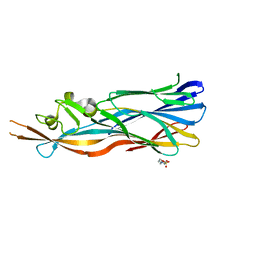
Structure of the Y250A mutant of the PANTON-VALENTINE LEUCOCIDIN S component from STAPHYLOCOCCUS AUREUS
Descriptor: 1,2-ETHANEDIOL, CITRATE ANION, LukS-PV
Authors:Maveyraud, L, Guerin, F, Laventie, B.J, Prevost, G, Mourey, L.
Deposit date:2013-01-28
Release date:2014-01-29
Last modified:2023-11-08
Method:X-RAY DIFFRACTION (1.55 Å)
Cite:Residues essential for panton-valentine leukocidin s component binding to its cell receptor suggest both plasticity and adaptability in its interaction surface
Plos One, 9, 2014
4IZL
DownloadVisualize
BU of 4izl by Molmil
Structure Of The N248A Mutant of the PANTON-VALENTINE LEUCOCIDIN S Component from STAPHYLOCOCCUS AUREUS
Descriptor: LukS-PV
Authors:Maveyraud, L, Laventie, B.J, Prevost, G, Mourey, L.
Deposit date:2013-01-30
Release date:2014-01-29
Last modified:2023-11-08
Method:X-RAY DIFFRACTION (2.8 Å)
Cite:Residues essential for panton-valentine leukocidin s component binding to its cell receptor suggest both plasticity and adaptability in its interaction surface
Plos One, 9, 2014

218500

PDB entries from 2024-04-17

PDB statisticsPDBj update infoContact PDBjnumon