Loading
PDBj
MenuPDBj@FacebookPDBj@TwitterPDBj@YouTubewwPDB FoundationwwPDB
RCSB PDBPDBeBMRBAdv. SearchSearch help
Search by PDB author
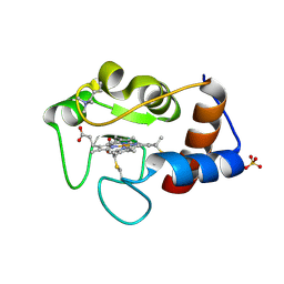
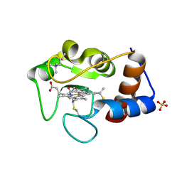
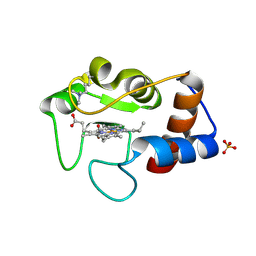
1CRH
DownloadVisualize
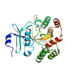
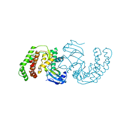
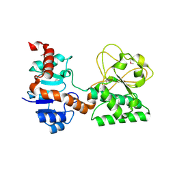
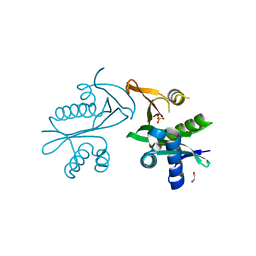
BU of 1crh by Molmil
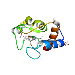
THE ROLE OF A CONSERVED INTERNAL WATER MOLECULE AND ITS ASSOCIATED HYDROGEN BOND NETWORK IN CYTOCHROME C
Descriptor: CYTOCHROME C, HEME C, SULFATE ION
Authors:Berghuis, A.M, Brayer, G.D.
Deposit date:1993-08-06
Release date:1994-01-31
Last modified:2021-03-03
Method:X-RAY DIFFRACTION (1.9 Å)
Cite:The role of a conserved internal water molecule and its associated hydrogen bond network in cytochrome c.
J.Mol.Biol., 236, 1994
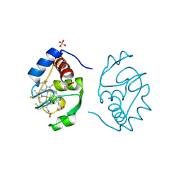
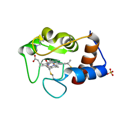
1CRI
DownloadVisualize
BU of 1cri by Molmil
THE ROLE OF A CONSERVED INTERNAL WATER MOLECULE AND ITS ASSOCIATED HYDROGEN BOND NETWORK IN CYTOCHROME C
Descriptor: CYTOCHROME C, PROTOPORPHYRIN IX CONTAINING FE, SULFATE ION
Authors:Berghuis, A.M, Brayer, G.D.
Deposit date:1993-08-06
Release date:1994-01-31
Last modified:2017-11-29
Method:X-RAY DIFFRACTION (2 Å)
Cite:The role of a conserved internal water molecule and its associated hydrogen bond network in cytochrome c.
J.Mol.Biol., 236, 1994
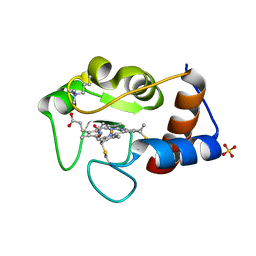
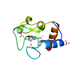
1CRG
DownloadVisualize
BU of 1crg by Molmil
THE ROLE OF A CONSERVED INTERNAL WATER MOLECULE AND ITS ASSOCIATED HYDROGEN BOND NETWORK IN CYTOCHROME C
Descriptor: CYTOCHROME C, HEME C, SULFATE ION
Authors:Berghuis, A.M, Brayer, G.D.
Deposit date:1993-08-06
Release date:1994-01-31
Last modified:2021-03-03
Method:X-RAY DIFFRACTION (2 Å)
Cite:The role of a conserved internal water molecule and its associated hydrogen bond network in cytochrome c.
J.Mol.Biol., 236, 1994
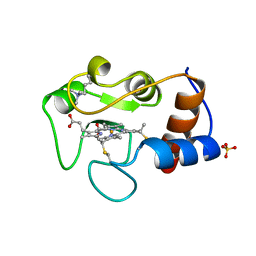
1CRJ
DownloadVisualize
BU of 1crj by Molmil
THE ROLE OF A CONSERVED INTERNAL WATER MOLECULE AND ITS ASSOCIATED HYDROGEN BOND NETWORK IN CYTOCHROME C
Descriptor: CYTOCHROME C, HEME C, SULFATE ION
Authors:Berghuis, A.M, Brayer, G.D.
Deposit date:1993-08-06
Release date:1994-01-31
Last modified:2021-03-03
Method:X-RAY DIFFRACTION (2.05 Å)
Cite:The role of a conserved internal water molecule and its associated hydrogen bond network in cytochrome c.
J.Mol.Biol., 236, 1994
2YCC
DownloadVisualize
BU of 2ycc by Molmil
OXIDATION STATE-DEPENDENT CONFORMATIONAL CHANGES IN CYTOCHROME C
Descriptor: CYTOCHROME C, HEME C, SULFATE ION
Authors:Berghuis, A.M, Brayer, G.D.
Deposit date:1991-01-29
Release date:1992-07-15
Last modified:2021-03-03
Method:X-RAY DIFFRACTION (1.9 Å)
Cite:Oxidation state-dependent conformational changes in cytochrome c.
J.Mol.Biol., 223, 1992
1GIT
DownloadVisualize
BU of 1git by Molmil
STRUCTURE OF GTP-BINDING PROTEIN
Descriptor: G PROTEIN GI ALPHA 1, GUANOSINE-5'-DIPHOSPHATE, PHOSPHATE ION
Authors:Berghuis, A.M, Lee, E, Sprang, S.R.
Deposit date:1996-10-16
Release date:1997-02-12
Last modified:2024-02-07
Method:X-RAY DIFFRACTION (2.6 Å)
Cite:Structure of the GDP-Pi complex of Gly203-->Ala gialpha1: a mimic of the ternary product complex of galpha-catalyzed GTP hydrolysis.
Structure, 4, 1996
1CTY
DownloadVisualize
BU of 1cty by Molmil
MUTATION OF TYROSINE-67 IN CYTOCHROME C SIGNIFICANTLY ALTERS THE LOCAL HEME ENVIRONMENT
Descriptor: CYTOCHROME C, HEME C, SULFATE ION
Authors:Berghuis, A.M, Brayer, G.D.
Deposit date:1993-02-15
Release date:1993-07-15
Last modified:2021-03-03
Method:X-RAY DIFFRACTION (2.2 Å)
Cite:Mutation of tyrosine-67 to phenylalanine in cytochrome c significantly alters the local heme environment.
J.Mol.Biol., 235, 1994
1CTZ
DownloadVisualize
BU of 1ctz by Molmil
MUTATION OF TYROSINE-67 IN CYTOCHROME C SIGNIFICANTLY ALTERS THE LOCAL HEME ENVIRONMENT
Descriptor: CYTOCHROME C, HEME C, SULFATE ION
Authors:Berghuis, A.M, Brayer, G.D.
Deposit date:1993-02-15
Release date:1993-07-15
Last modified:2021-03-03
Method:X-RAY DIFFRACTION (1.9 Å)
Cite:Mutation of tyrosine-67 to phenylalanine in cytochrome c significantly alters the local heme environment.
J.Mol.Biol., 235, 1994
1IRV
DownloadVisualize
BU of 1irv by Molmil
CYTOCHROME C ISOZYME 1, REDUCED, MUTANT WITH ILE 75 REPLACED BY MET AND CYS 102 REPLACED BY THR
Descriptor: CYTOCHROME C, PROTOPORPHYRIN IX CONTAINING FE, SULFATE ION
Authors:Berghuis, A.M, Brayer, G.D.
Deposit date:1996-06-27
Release date:1997-01-11
Last modified:2021-11-03
Method:X-RAY DIFFRACTION (1.9 Å)
Cite:Mechanistic and structural contributions of critical surface and internal residues to cytochrome c electron transfer reactivity.
Biochemistry, 35, 1996
1IRW
DownloadVisualize
BU of 1irw by Molmil
CYTOCHROME C ISOZYME 1, REDUCED, MUTANT WITH ASN 52 REPLACED BY ALA AND CYS 102 REPLACED BY THR
Descriptor: CYTOCHROME C, PROTOPORPHYRIN IX CONTAINING FE, SULFATE ION
Authors:Berghuis, A.M, Brayer, G.D.
Deposit date:1996-06-27
Release date:1997-01-11
Last modified:2021-11-03
Method:X-RAY DIFFRACTION (2 Å)
Cite:Mechanistic and structural contributions of critical surface and internal residues to cytochrome c electron transfer reactivity.
Biochemistry, 35, 1996
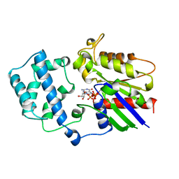
3TM0
DownloadVisualize
BU of 3tm0 by Molmil
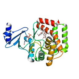
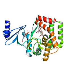
Crystal Structure of 3',5"-Aminoglycoside Phosphotransferase Type IIIa AMPPNP Butirosin A Complex
Descriptor: (2S)-4-amino-N-[(1R,2S,3R,4R,5S)-5-amino-4-[(2,6-diamino-2,6-dideoxy-alpha-D-glucopyranosyl)oxy]-2-hydroxy-3-(beta-D-xylofuranosyloxy)cyclohexyl]-2-hydroxybutanamide, Aminoglycoside 3'-phosphotransferase, MAGNESIUM ION, ...
Authors:Berghuis, A.M, Fong, D.H.
Deposit date:2011-08-30
Release date:2011-10-19
Last modified:2023-09-13
Method:X-RAY DIFFRACTION (2.1 Å)
Cite:Structural basis of APH(3')-IIIa-mediated resistance to N1-substituted aminoglycoside antibiotics.
Antimicrob.Agents Chemother., 53, 2009
3Q2M
DownloadVisualize
BU of 3q2m by Molmil
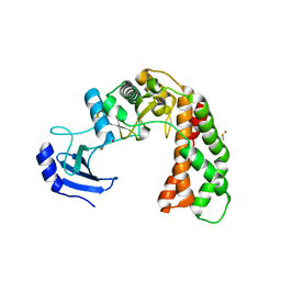
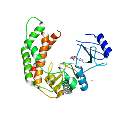
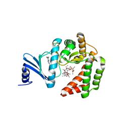
Crystal Structure of Spectinomycin Phosphotransferase, APH(9)-Ia, Protein Kinase Inhibitor CKI-7 Complex
Descriptor: N-(2-AMINOETHYL)-5-CHLOROISOQUINOLINE-8-SULFONAMIDE, NICKEL (II) ION, Spectinomycin phosphotransferase
Authors:Berghuis, A.M, Fong, D.H, Xiong, B, Hwang, J.
Deposit date:2010-12-20
Release date:2011-05-18
Last modified:2023-09-13
Method:X-RAY DIFFRACTION (2.9 Å)
Cite:Crystal structures of two aminoglycoside kinases bound with a eukaryotic protein kinase inhibitor.
Plos One, 6, 2011
2Q99
DownloadVisualize
BU of 2q99 by Molmil
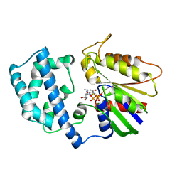
Crystal Structure of Saccharopine Dehydrogenase from Saccharomyces cerevisiae
Descriptor: Saccharopine dehydrogenase [NAD+, L-lysine-forming
Authors:Berghuis, A.M, Burk, D.L.
Deposit date:2007-06-12
Release date:2007-11-06
Last modified:2017-10-18
Method:X-RAY DIFFRACTION (1.64 Å)
Cite:Structural Studies of the Final Enzyme in the alpha-Aminoadipate Pathway-Saccharopine Dehydrogenase from Saccharomyces cerevisiae
J.Mol.Biol., 373, 2007
5E96
DownloadVisualize
BU of 5e96 by Molmil
Crystal structure of aminoglycoside 6'-acetyltransferase type Ii
Descriptor: 1,2-ETHANEDIOL, Aminoglycoside 6'-acetyltransferase, PHOSPHATE ION
Authors:Berghuis, A.M, Burk, D.L, Baettig, O.M, Shi, K.
Deposit date:2015-10-14
Release date:2016-07-06
Last modified:2023-09-27
Method:X-RAY DIFFRACTION (2.1 Å)
Cite:Comprehensive characterization of ligand-induced plasticity changes in a dimeric enzyme.
Febs J., 283, 2016
3I1A
DownloadVisualize
BU of 3i1a by Molmil
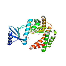
Crystal Structure of apo Spectinomycin Phosphotransferase, APH(9)-Ia
Descriptor: 2-(N-MORPHOLINO)-ETHANESULFONIC ACID, DIMETHYL SULFOXIDE, Spectinomycin phosphotransferase, ...
Authors:Berghuis, A.M, Fong, D.H, Lemke, C.T, Hwang, J, Xiong, B.
Deposit date:2009-06-25
Release date:2010-01-19
Last modified:2024-02-21
Method:X-RAY DIFFRACTION (1.7 Å)
Cite:Structure of the antibiotic resistance factor spectinomycin phosphotransferase from Legionella pneumophila.
J.Biol.Chem., 285, 2010
3I0Q
DownloadVisualize
BU of 3i0q by Molmil
Crystal Structure of the AMP-bound complex of Spectinomycin Phosphotransferase, APH(9)-Ia
Descriptor: ADENOSINE MONOPHOSPHATE, NICKEL (II) ION, Spectinomycin phosphotransferase
Authors:Berghuis, A.M, Fong, D.H, Lemke, C.T, Hwang, J.-Y, Xiong, B.
Deposit date:2009-06-25
Release date:2010-01-19
Last modified:2023-09-06
Method:X-RAY DIFFRACTION (2.8 Å)
Cite:Structure of the antibiotic resistance factor spectinomycin phosphotransferase from Legionella pneumophila.
J.Biol.Chem., 285, 2010
1GIA
DownloadVisualize
BU of 1gia by Molmil
STRUCTURE OF ACTIVE CONFORMATIONS OF GIA1 AND THE MECHANISM OF GTP HYDROLYSIS
Descriptor: 5'-GUANOSINE-DIPHOSPHATE-MONOTHIOPHOSPHATE, G PROTEIN GI ALPHA 1, MAGNESIUM ION
Authors:Berghuis, A.M, Coleman, D.E, Sprang, S.R.
Deposit date:1994-07-20
Release date:1994-09-30
Last modified:2024-02-07
Method:X-RAY DIFFRACTION (2 Å)
Cite:Structures of active conformations of Gi alpha 1 and the mechanism of GTP hydrolysis.
Science, 265, 1994
5IGH
DownloadVisualize
BU of 5igh by Molmil
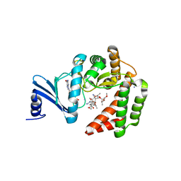
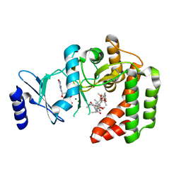
Macrolide 2'-phosphotransferase type I
Descriptor: Macrolide 2'-phosphotransferase, SULFATE ION
Authors:Berghuis, A.M, Fong, D.H.
Deposit date:2016-02-28
Release date:2017-04-26
Last modified:2020-01-08
Method:X-RAY DIFFRACTION (1.55 Å)
Cite:Structural Basis for Kinase-Mediated Macrolide Antibiotic Resistance.
Structure, 25, 2017
5IGV
DownloadVisualize
BU of 5igv by Molmil
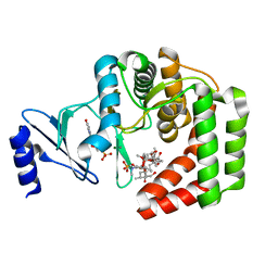
Macrolide 2'-phosphotransferase type II - complex with GDP and azithromycin
Descriptor: AZITHROMYCIN, CALCIUM ION, GUANOSINE-5'-DIPHOSPHATE, ...
Authors:Berghuis, A.M, Fong, D.H.
Deposit date:2016-02-28
Release date:2017-04-26
Last modified:2023-09-27
Method:X-RAY DIFFRACTION (1.55 Å)
Cite:Structural Basis for Kinase-Mediated Macrolide Antibiotic Resistance.
Structure, 25, 2017
5IGR
DownloadVisualize
BU of 5igr by Molmil
Macrolide 2'-phosphotransferase type I - complex with GDP and oleandomycin
Descriptor: (3S,5R,6S,7R,8R,11R,12S,13R,14S,15S)-6-HYDROXY-5,7,8,11,13,15-HEXAMETHYL-4,10-DIOXO-14-{[3,4,6-TRIDEOXY-3-(DIMETHYLAMINO)-BETA-D-XYLO-HEXOPYRANOSYL]OXY}-1,9-DIOXASPIRO[2.13]HEXADEC-12-YL 2,6-DIDEOXY-3-O-METHYL-ALPHA-L-ARABINO-HEXOPYRANOSIDE, AMMONIUM ION, GUANOSINE-5'-DIPHOSPHATE, ...
Authors:Berghuis, A.M, Fong, D.H.
Deposit date:2016-02-28
Release date:2017-04-26
Last modified:2024-03-06
Method:X-RAY DIFFRACTION (1.6 Å)
Cite:Structural Basis for Kinase-Mediated Macrolide Antibiotic Resistance.
Structure, 25, 2017
5IH1
DownloadVisualize
BU of 5ih1 by Molmil
Macrolide 2'-phosphotransferase type II - complex with GDP and phosphorylated josamycin
Descriptor: CALCIUM ION, GUANOSINE-5'-DIPHOSPHATE, MAGNESIUM ION, ...
Authors:Berghuis, A.M, Fong, D.H.
Deposit date:2016-02-28
Release date:2017-04-26
Last modified:2024-03-06
Method:X-RAY DIFFRACTION (1.31 Å)
Cite:Structural Basis for Kinase-Mediated Macrolide Antibiotic Resistance.
Structure, 25, 2017
5IGU
DownloadVisualize
BU of 5igu by Molmil
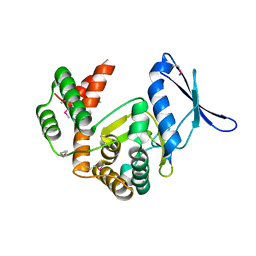
Macrolide 2'-phosphotransferase type II
Descriptor: Macrolide 2'-phosphotransferase II
Authors:Berghuis, A.M, Fong, D.H.
Deposit date:2016-02-28
Release date:2017-04-26
Last modified:2020-01-08
Method:X-RAY DIFFRACTION (2.1 Å)
Cite:Structural Basis for Kinase-Mediated Macrolide Antibiotic Resistance.
Structure, 25, 2017
5IH0
DownloadVisualize
BU of 5ih0 by Molmil
Macrolide 2'-phosphotransferase type II Y92M mutant - complex with GDP and erythromycin
Descriptor: ACETATE ION, ERYTHROMYCIN A, GUANOSINE-5'-DIPHOSPHATE, ...
Authors:Berghuis, A.M, Fong, D.H.
Deposit date:2016-02-28
Release date:2017-04-26
Last modified:2023-09-27
Method:X-RAY DIFFRACTION (1.65 Å)
Cite:Structural Basis for Kinase-Mediated Macrolide Antibiotic Resistance.
Structure, 25, 2017
5IGJ
DownloadVisualize
BU of 5igj by Molmil
Macrolide 2'-phosphotransferase type I - complex with guanosine and clarithromycin
Descriptor: CLARITHROMYCIN, GUANOSINE, ISOPROPYL ALCOHOL, ...
Authors:Berghuis, A.M, Fong, D.H.
Deposit date:2016-02-28
Release date:2017-04-26
Last modified:2023-09-27
Method:X-RAY DIFFRACTION (1.4 Å)
Cite:Structural Basis for Kinase-Mediated Macrolide Antibiotic Resistance.
Structure, 25, 2017
5IGW
DownloadVisualize
BU of 5igw by Molmil
Macrolide 2'-phosphotransferase type II - complex with GDP and clarithromycin
Descriptor: CLARITHROMYCIN, GUANOSINE-5'-DIPHOSPHATE, MAGNESIUM ION, ...
Authors:Berghuis, A.M, Fong, D.H.
Deposit date:2016-02-28
Release date:2017-04-26
Last modified:2023-09-27
Method:X-RAY DIFFRACTION (2.096 Å)
Cite:Structural Basis for Kinase-Mediated Macrolide Antibiotic Resistance.
Structure, 25, 2017

218853

PDB entries from 2024-04-24

PDB statisticsPDBj update infoContact PDBjnumon